Clear Sky Science · en
Label-free in vivo molecular profiling of the human retina by non-resonant Raman spectroscopy
Seeing Tiny Changes Before Vision Fades
Many eye and brain diseases quietly damage nerve cells long before people notice blurry vision or memory loss. By the time standard eye scans reveal visible harm, much of the damage is already permanent. This study explores a light-based technique that can read the chemical makeup of living tissue inside the eye without any dyes or injections. The goal is to spot very early warning signs of disease in the retina—the thin nerve layer at the back of the eye that also serves as a convenient "window" into the brain.
A Gentle Light That Listens to Molecules
The researchers rely on a method called Raman spectroscopy, which uses a safe, narrow beam of laser light. When this light hits tissue, most of it bounces back unchanged, but a tiny fraction interacts with the molecules and returns slightly shifted in color. These shifts form a kind of barcode that reflects the presence of broad chemical groups such as fats, proteins, sugars and DNA building blocks. Because it does not require contrast dyes or physical contact, this approach is naturally gentle and label-free, making it attractive for repeated measurements in people.
Finding the Quiet Corner of the Retina
Earlier attempts to use this technique in the living eye struggled with a major obstacle: strong natural glow from pigments in the central retina that drowns out the subtle molecular signal. In this work, the team carefully scanned many positions across the back of the eye in a volunteer while using conventional imaging to guide the laser spot. They discovered that almost all regions produced overwhelming background light, except for one key area: the optic nerve head, where nerve fibers from the retina bundle together and exit the eye. Here, the troublesome pigments are naturally absent, allowing the molecular signal to emerge clearly and revealing features linked to fats, proteins, sugars and genetic material. 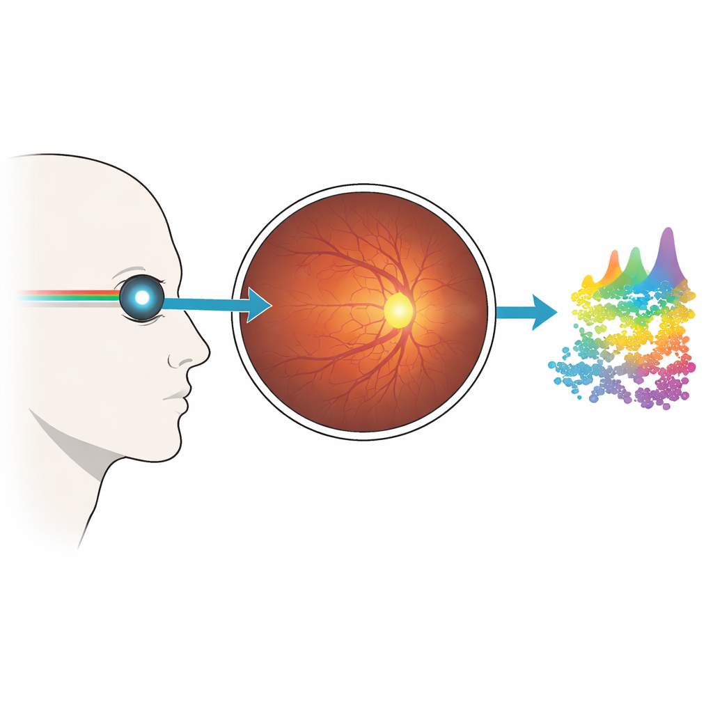
Watching an Individual Eye Over Time
Once they identified this sweet spot, the scientists repeatedly measured the same region in one person over seven sessions spanning four months. They used advanced data cleaning methods to remove noisy readings and to correct for small changes in overall brightness. The resulting molecular fingerprints were highly consistent from visit to visit, confirming that the method is stable enough for practical use. At the same time, some portions of the signal showed real variation, hinting at changes in components such as certain fats, sugars and amino acids that may reflect the natural ebb and flow of nerve cell activity and support cells in this busy crossroads of the visual pathway.
Tracing the Chemical Footprint of Aging
To find out whether this light-based fingerprint can track how the eye’s nerve tissue changes with age, the team then examined 21 healthy volunteers ranging from their twenties to late seventies. For each person, they recorded several spectra at the optic nerve head and compared three age groups: younger than 45, middle-aged, and older than 65. Statistical analysis showed that the spectra clustered differently by age, even though all participants were clinically normal. In particular, signals linked mainly to certain fats—such as cholesterol-like molecules and components of cell membranes—tended to grow stronger with age, while bands influenced by proteins and DNA-related structures tended to decline. Together, these shifts point to a gradual remodeling of the nerve tissue’s chemical landscape as people get older. 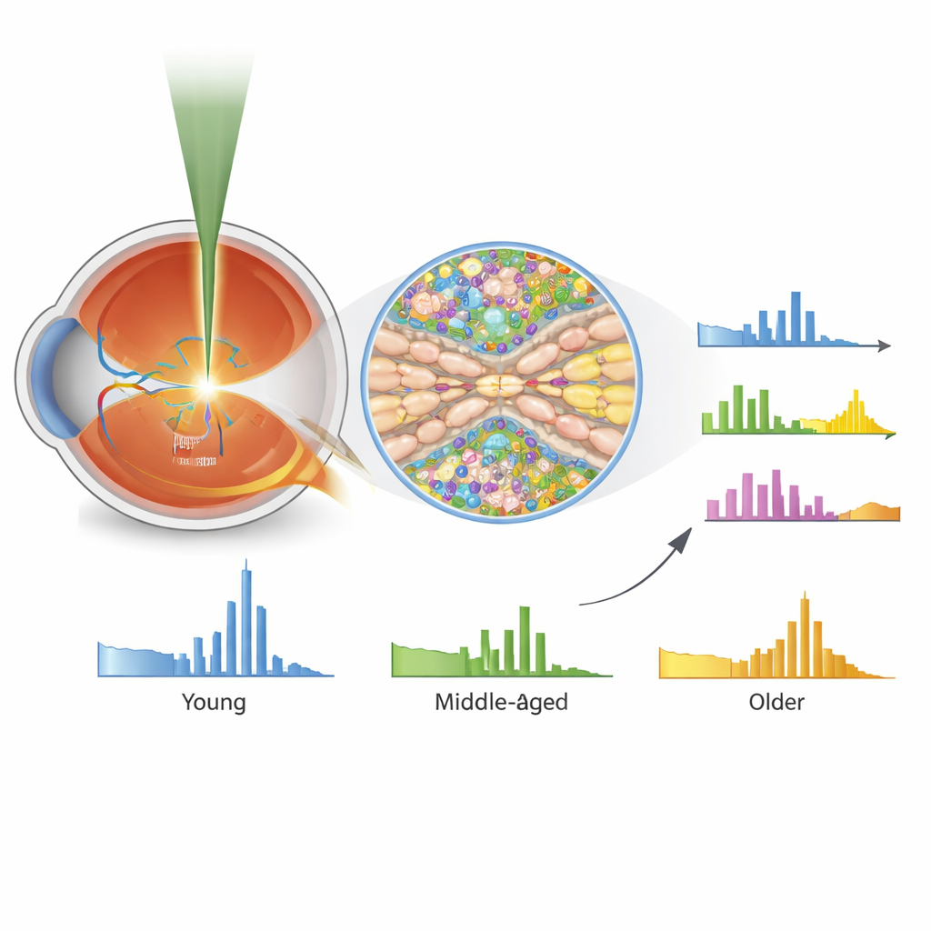
What This Could Mean for Eye and Brain Health
By showing that clear, repeatable molecular fingerprints can be measured safely from the optic nerve head in living people, this study lays the groundwork for a new kind of eye exam. Instead of waiting for visible thinning of nerve layers, doctors might one day monitor subtle chemical changes that appear earlier in conditions such as glaucoma, age-related macular degeneration, or even brain disorders that leave traces in the eye. The authors also caution that aging alone already causes measurable shifts in the retinal chemistry, so future work will need to carefully separate normal aging patterns from true disease signals. Nonetheless, this label-free technique offers a promising route toward earlier, more precise detection of nerve damage in both the eye and the brain.
Citation: Sentosa, R., Kendrisic, M., Salas, M. et al. Label-free in vivo molecular profiling of the human retina by non-resonant Raman spectroscopy. Commun Biol 9, 511 (2026). https://doi.org/10.1038/s42003-026-09744-2
Keywords: retina, Raman spectroscopy, optic nerve head, molecular imaging, aging