Clear Sky Science · en
Early life adversity impairs visually evoked innate defensive behaviors via oxytocin signaling
Why early hardship and quick reactions to danger matter
When something suddenly swoops toward us—a falling object, a fast car, a looming shadow—our brains are wired to trigger instant, automatic defenses. These split‑second reactions help keep us alive. Yet people who endured serious hardship in childhood are more likely to suffer accidental injuries later in life, hinting that their basic danger‑detection systems may be off. This study in mice asks a deceptively simple question: can early social adversity blunt an animal’s built‑in visual alarm system, and if so, what changes in the brain and body’s chemistry might explain it?
From stressful childhoods to slower escapes
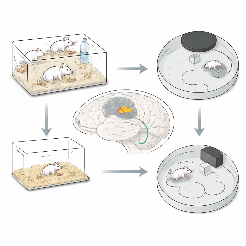
The researchers created a mouse model of early life adversity by briefly separating pups from their mothers and littermates each day. One group experienced this social deprivation very early, from birth to day 12; another group experienced it a little later, from day 10 to 20, a period when the brain is rapidly maturing. In adulthood, the mice were tested with a classic “looming” threat: a black disk rapidly expanding on a screen overhead, mimicking an approaching predator. Healthy mice instinctively sprint to a nearby shelter. Mice that had been separated during the later window, but not the earlier one, reacted more slowly, ran along longer and less direct paths, and spent less time safely tucked in the refuge afterward. Their bodies and general movement looked normal, and their anxiety levels in other tests were unchanged, suggesting that a specific defensive reflex—rather than overall health or mood—had been weakened.
A chemical signal in the midbrain spotlight
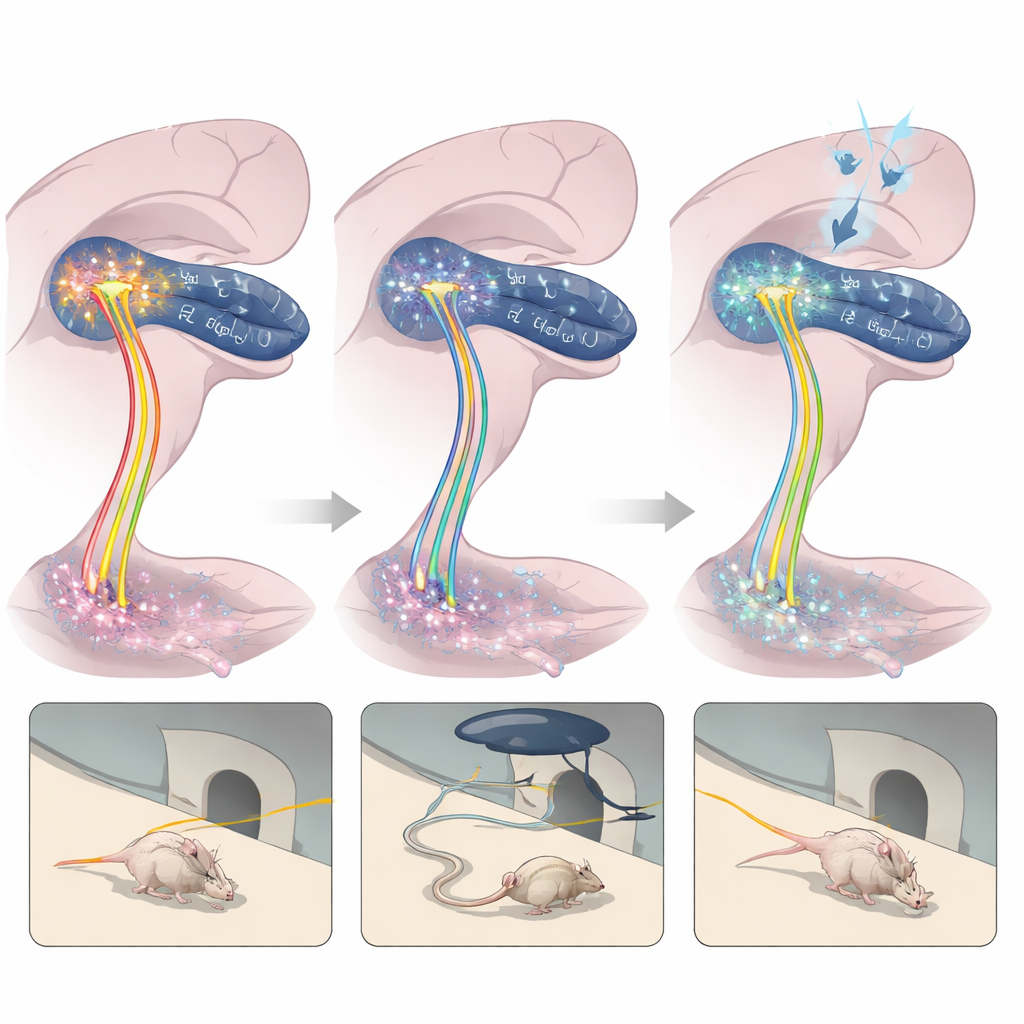
To understand why these mice were slower to flee, the team focused on oxytocin, a hormone best known for roles in bonding and social behavior but increasingly linked to fear and stress. Earlier human and animal work shows that early adversity often dampens oxytocin signaling. Here, the scientists examined a midbrain structure called the superior colliculus, which helps turn fast visual cues into rapid actions. Within its intermediate and deep layers, they measured the gene activity for the oxytocin receptor—the docking site that allows oxytocin to influence local nerve cells. Mice that had undergone later social deprivation showed fewer active cells and weaker oxytocin receptor signals in these layers after the looming test, even though the overall number of oxytocin‑producing neurons and the amount of oxytocin released elsewhere in the brain appeared unchanged. In other words, the problem seemed to lie at the receiving end of the signal.
Recreating and reversing the deficit
To test whether this weakened oxytocin reception actually caused the poorer defenses, the researchers used genetic tools to selectively reduce oxytocin receptors in the superior colliculus of otherwise normal adult mice. These animals then behaved much like the deprived group: they hesitated longer before reaching shelter, moved more slowly, and lingered outside the refuge. In a complementary experiment, the team turned on oxytocin‑carrying nerve fibers that run from a region deep in the brain, the paraventricular nucleus of the hypothalamus, down to the same midbrain layers. Light‑based stimulation of these fibers during looming trials sharpened the mice’s defensive responses—they darted to safety more quickly and stayed there longer. Together, these manipulations show that oxytocin‑sensitive circuits in this visual‑motor hub are both necessary and sufficient to tune the speed and strength of innate escape behaviors.
A potential path toward repair
Because giving oxytocin through the nose can deliver it to the brain and boost receptor activity, the scientists asked whether this simple treatment could offset the damage of early adversity. Adult mice that had experienced late social deprivation received intranasal oxytocin every other day for nearly two weeks before being exposed to the looming threat. Compared with deprived mice given only saline, treated animals ran faster toward the refuge and spent more time inside after the simulated attack, indicating a partial rescue of their defensive reflex. Although the study did not directly measure receptor levels after treatment, the behavioral improvement suggests that strengthening oxytocin signaling along the hypothalamus–midbrain pathway can restore at least some of the lost sensitivity to visual danger.
What this means for human health
These findings reveal a concrete chain linking early social hardship to altered brain chemistry and, ultimately, to dulled automatic reactions to visual threats. In mice, a brief but crucial window of maternal care appears to be especially important for building oxytocin‑sensitive circuits that transform a looming shadow into a rapid dash for safety. When that window is disturbed, the brain’s danger‑processing network becomes less responsive—but targeted boosting of oxytocin signaling can partly repair the deficit. While more work is needed to translate these insights to humans, the study offers a mechanistic framework for understanding how childhood adversity might quietly reshape our most basic survival responses, and it points to oxytocin pathways as promising candidates for future prevention and treatment strategies.
Citation: Tan, H., Su, J., Ma, S. et al. Early life adversity impairs visually evoked innate defensive behaviors via oxytocin signaling. Commun Biol 9, 467 (2026). https://doi.org/10.1038/s42003-026-09738-0
Keywords: early life adversity, oxytocin, innate fear, superior colliculus, mouse behavior