Clear Sky Science · en
Multidimensional data analysis and classification using SMIAL
Making Sense of Complex Scientific Pictures
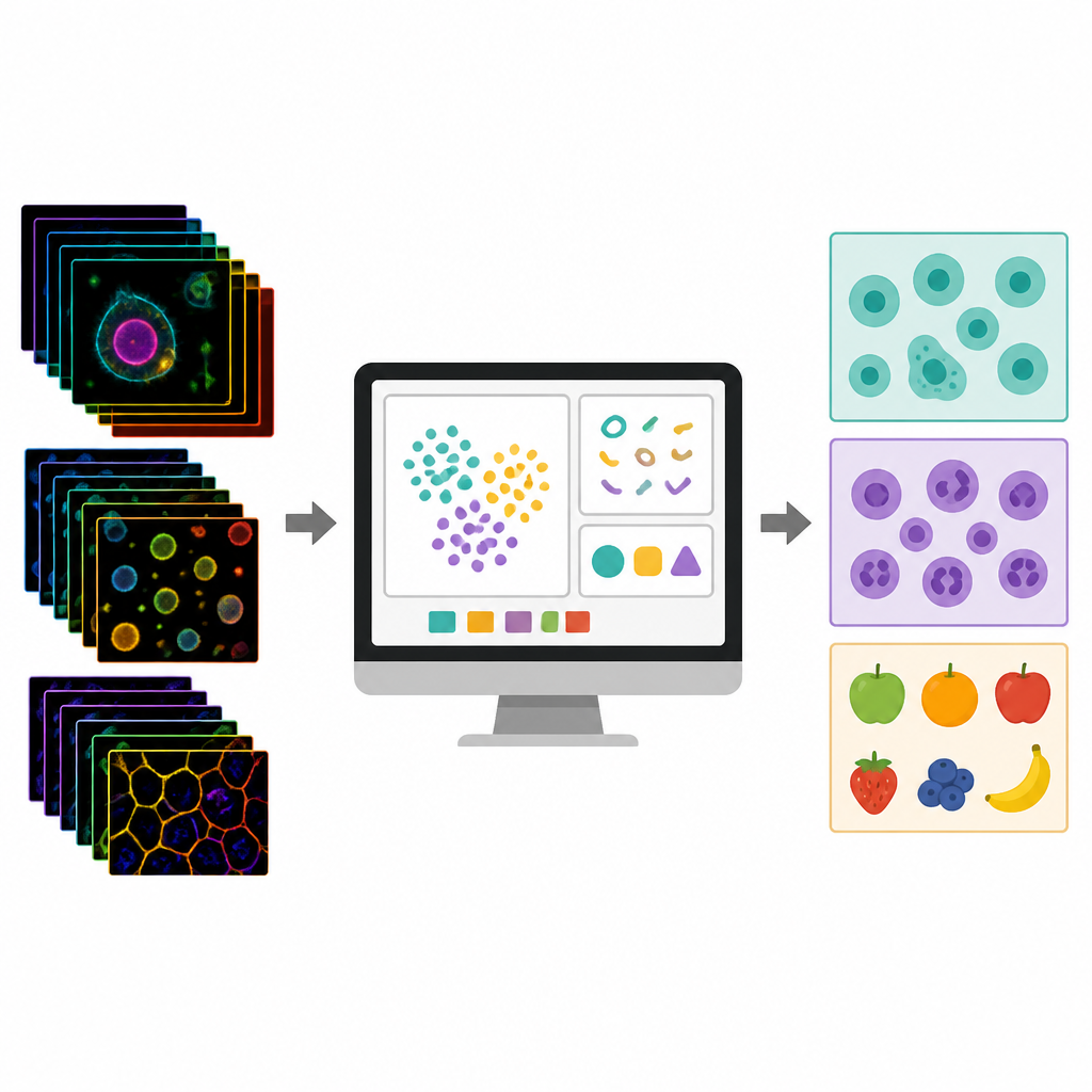
Modern microscopes can capture incredibly rich pictures of cells, tissues and even food, recording how they glow, reflect or change over time without adding any dyes or labels. These images hold clues about diseases, drug effects and product quality, but are often so complex that only experts with advanced programming skills can analyze them. This paper introduces SMIAL, a free software tool that helps scientists and clinicians turn these dense image collections into clear, trustworthy results using machine learning, all through a user friendly interface.
A Single Place for the Whole Journey
SMIAL is designed as a one stop workspace that guides users through the complete journey from raw images to final decisions. Instead of juggling several programs, users move through six panels that mirror how scientists typically work: loading images, outlining objects of interest, cleaning the data, measuring many properties and finally building and checking prediction models. The software accepts a wide range of inputs, including colored microscope images over many wavelengths, time lapse movies, simple measurement tables and even ready made models. At every step it saves the chosen settings so that others can repeat the work and understand exactly how the results were produced.
Teaching the Computer to Spot Melanoma Cells
To show what SMIAL can do, the authors first used it to distinguish between two types of melanoma cells and normal skin cells, using only their natural glow under different colors of light. They loaded image stacks with 29 spectral channels, aligned hand drawn cell outlines, and improved image quality with noise reduction, background removal and careful brightness calibration. SMIAL then measured more than a thousand traits per cell, capturing how bright each channel was, how textures varied and what the cell shapes looked like. A data cleaning step automatically removed weak or overlapping traits, shrinking the list to a few dozen informative ones. With these, a simple logistic regression classifier correctly separated the three cell types with high accuracy, as confirmed by strong performance scores and clear clusters of cells in summary plots.
Watching Cells Respond to a Drug Over Time
In a second case study, SMIAL tracked how retinal cells responded over days to rotenone, a chemical that stresses mitochondria, the energy producers of the cell. The team examined natural mitochondrial glow at several time points in treated and untreated cells. After testing different noise filters, they kept the one that preserved fine network details. SMIAL measured intensity, shape and texture of the mitochondrial web and also computed how these measures changed between time points. Treated mitochondria became brighter, more fragmented and less circular over 72 hours. When time based features were added to the model, the software could better distinguish treated from control cells than when relying on static snapshots alone, highlighting the power of following changes instead of single images.
Judging Fruit Ripeness Without Cutting It Open
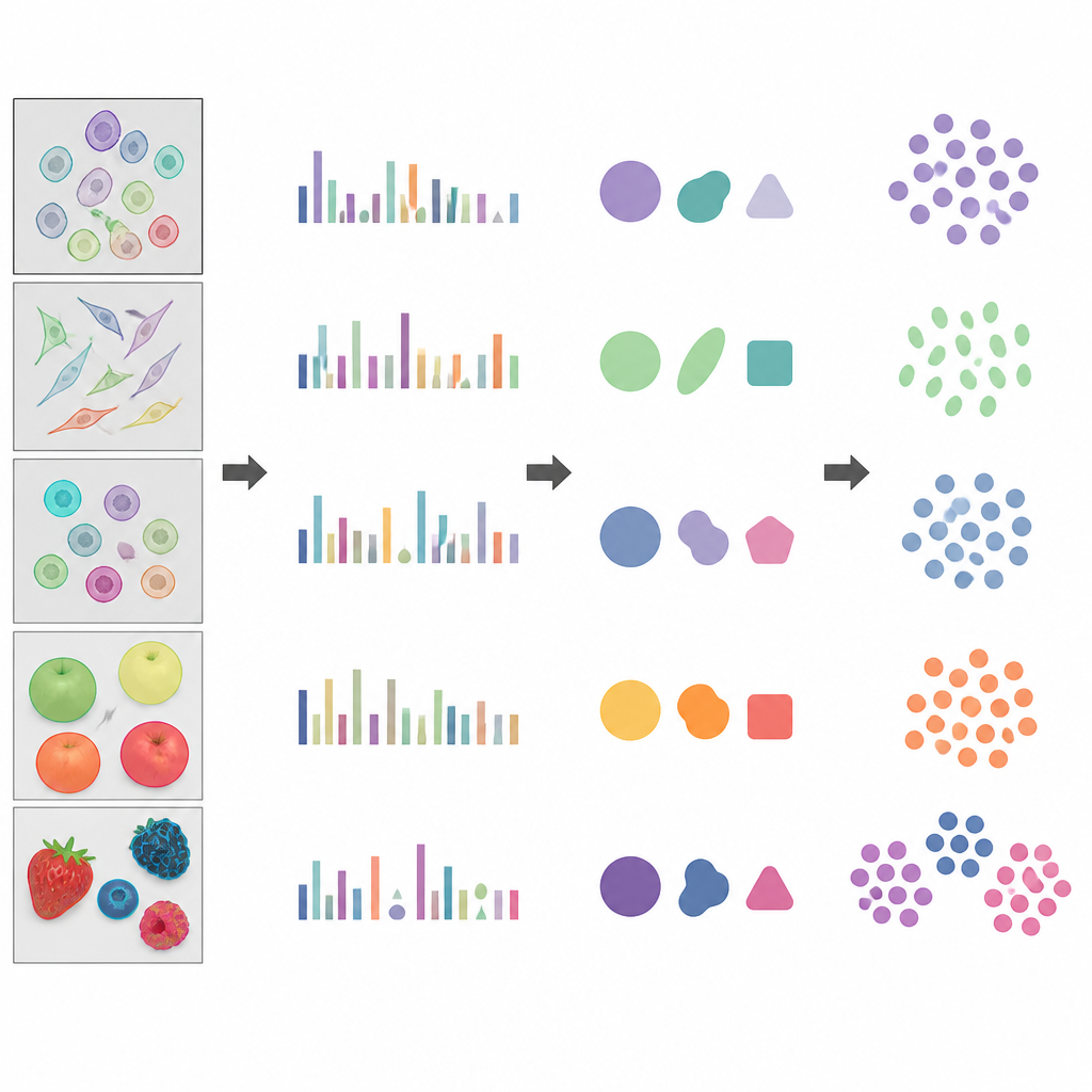
The third example turned to food quality, using public hyperspectral images of fruit at unripe, ripe and overripe stages, each recorded in 224 wavelengths. SMIAL automatically outlined each fruit based on contrast with the background and achieved segmentation quality close to careful manual drawings. It then extracted thousands of features describing brightness, shape and texture across wavelengths. While single measurements did not cleanly separate ripeness levels, SMIAL combined feature selection, clever handling of uneven class sizes and a support vector machine classifier to improve recognition, particularly for ripe fruit. This demonstrates that the same tool can handle both medical style datasets and industrial quality control tasks.
Lowering the Barrier to Smart Image Analysis
Overall, the study shows that SMIAL can turn large, complex image collections into reliable classification models for very different problems, from cancer cell detection to drug response and fruit ripeness. By wrapping many advanced analysis steps in a clear graphical interface and preserving full records of the chosen settings, it allows researchers who are not programming experts to build, share and repeat sophisticated machine learning workflows. In practical terms, this means more labs can extract useful insights from label free imaging, helping to speed progress in diagnostics, treatment monitoring and non invasive quality testing.
Citation: Knab, A., Handley, S., Xu, X. et al. Multidimensional data analysis and classification using SMIAL. Commun Biol 9, 650 (2026). https://doi.org/10.1038/s42003-026-09630-x
Keywords: label free imaging, multispectral microscopy, machine learning software, image based classification, hyperspectral imaging