Clear Sky Science · en
Generalizable CT-free PET attenuation and scatter correction via few-shot cross domain adaptation
Why this matters for patients and doctors
Positron emission tomography (PET) scans help doctors see how cancers and other diseases behave inside the body. But to get accurate PET images, patients usually need an extra computed tomography (CT) scan, which adds radiation and can sometimes misalign with the PET images. This study introduces a way to clean up PET images without relying on CT, using artificial intelligence that can quickly adapt to new hospitals and scanner types with only a handful of patient examples.
The problem of clearer pictures with less radiation
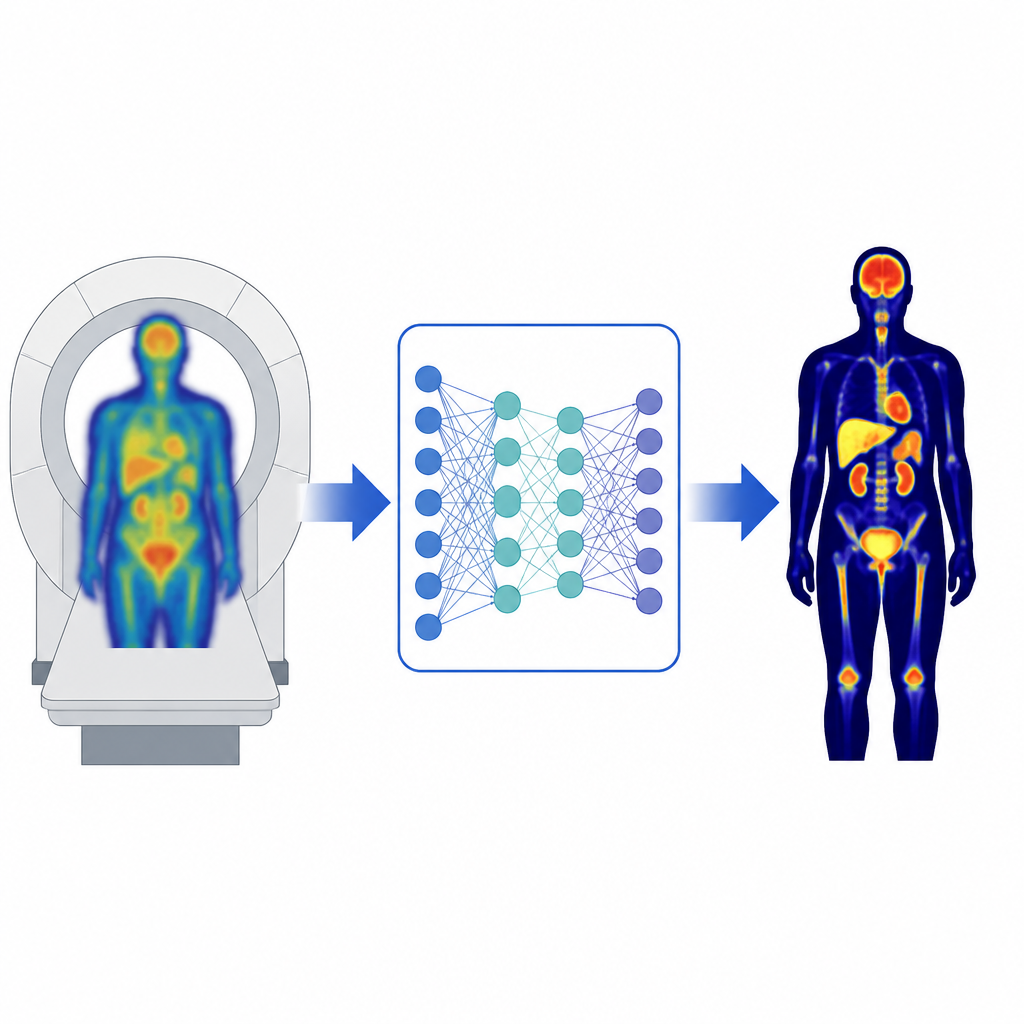
Modern PET/CT scanners combine two kinds of imaging: PET, which shows how tissues use energy or specific molecules, and CT, which shows body structure. The CT part helps correct for signal loss and scattering in PET so that doctors can trust the numbers they see in tumors and organs. However, the CT scan can contribute up to half of the total radiation dose in a whole-body PET/CT exam, which is especially concerning for children and other sensitive groups. Even when very low dose CT is used, natural breathing and patient motion can cause the CT and PET images to fall out of step, creating streaks and false hot or cold spots. Researchers therefore want methods that correct PET images without needing CT at all.
How artificial intelligence steps in
Recent deep learning methods try to fix uncorrected PET images directly. Some learn to predict a virtual CT image, while others learn to convert a blurry, uncorrected PET image into a sharper, corrected one in a single step. These systems can work well where they were trained, but they often struggle when the hospital changes scanners, tracers, or patient populations. Small differences in machines, settings, or the radioactive tracers used to highlight disease can shift the appearance of PET images and confuse a model that has only seen a narrow set of examples. The authors argue that it is unrealistic to train a single, fixed model that will always work everywhere, and that PET correction should instead be treated as a living system that can adapt to each new clinical setting.
A flexible PET correction model that learns from a few cases
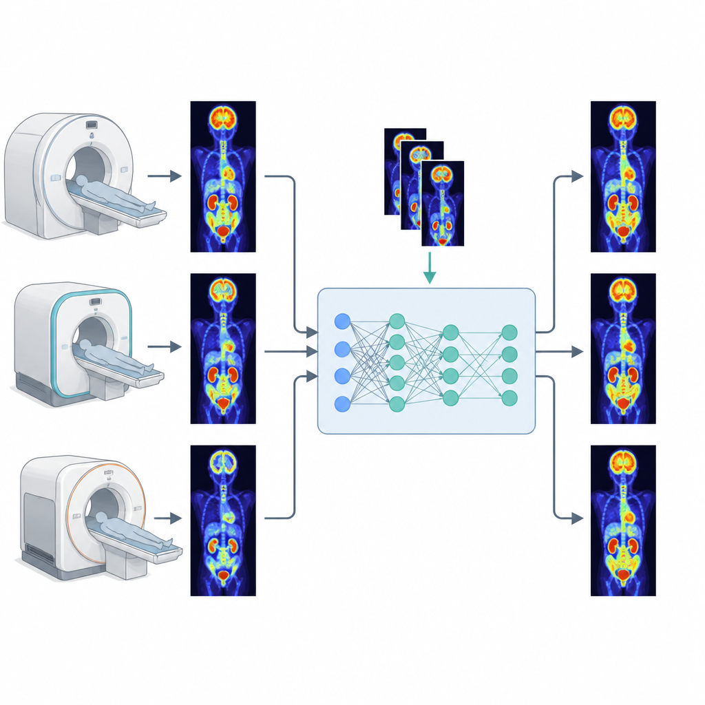
The team developed a framework called CrossPET-Adapt that uses a two-stage learning process. First, they trained a deep network on a very large set of PET scans from one widely used tracer, 18F-FDG, collected at a major center with a long axial field of view scanner. Rather than directly drawing a new PET image, the network learns a smooth correction map that scales each pixel of the input, reflecting how real physical losses in PET behave like changes in signal strength. It also uses simple statistics from each input scan, such as average brightness and variation, to tune its internal features so it can recognize how different scanners and tracers shift image appearance. In the second stage, the model is fine-tuned on just one to five patients from a new domain, such as a different tracer, scanner, or hospital, allowing it to capture local quirks without retraining from scratch.
Testing across tracers, scanners, and hospitals
To see how well this idea works, the researchers assembled a dataset of 1539 patients from 11 groups, including four tracers, several scanner models from different vendors, and three outside hospitals. They compared three strategies: models trained on a single tracer, a joint model trained on mixed tracers, and their few-shot adaptation approach. In settings where the tracers matched the training data, the mixed model performed slightly better, as expected. But in new environments with unseen tracers, scanners, or centers, the adapted model consistently delivered higher image similarity, lower error, and more reliable standardized uptake values, even when it was fine-tuned on only one or a few subjects. In lymphoma patients, the deep learning corrected images closely matched standard PET/CT results, sharply reducing errors in tumor volume and uptake measures that guide treatment decisions.
What this means for future PET scans
The study shows that CT-free PET correction can be both practical and reliable when paired with a few-shot adaptation strategy. Instead of collecting hundreds of labeled scans every time a hospital installs a new scanner or starts using a new tracer, clinicians could fine-tune a shared base model with just a handful of local cases in under ten minutes on a single graphics card. This approach could lower radiation exposure, simplify workflows, and make advanced PET imaging more consistent across sites, while still preserving the quantitative detail that oncologists and other specialists depend on.
Citation: Wang, H., Wen, M., Qiao, X. et al. Generalizable CT-free PET attenuation and scatter correction via few-shot cross domain adaptation. npj Digit. Med. 9, 374 (2026). https://doi.org/10.1038/s41746-026-02760-w
Keywords: PET imaging, attenuation correction, deep learning, domain adaptation, medical imaging