Clear Sky Science · en
A unified deep learning framework for cross-platform harmonization of multi-tracer PET quantification in neurodegenerative disease
Why this matters for brain scans
Brain scans that track disease markers are becoming central to diagnosing and monitoring conditions like Alzheimer’s disease, but the numbers doctors rely on can shift simply because a patient is scanned on a different machine. This study introduces a deep learning approach that makes measurements from low‑radiation PET‑MRI scans line up closely with traditional PET‑CT scans, so that thresholds and follow‑up comparisons stay meaningful even when patients move between hospitals or scanners.
The challenge of mixed scan technologies
Positron emission tomography (PET) helps doctors see abnormal proteins and brain activity linked to dementia and Parkinson’s disease. Many clinics now use hybrid PET‑MRI scanners, which cut radiation exposure compared with PET‑CT and can be preferable for people who need repeated scans. However, PET‑MRI and PET‑CT measure tracer uptake differently, largely because MRI has to guess how tissues absorb radiation while CT measures it directly. This mismatch can lead to 10–25% differences in key values between machines, enough to push a patient just above or below a treatment cutoff even when their brain biology has not changed.
A deep learning bridge between machines
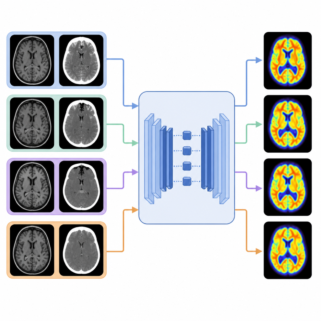
The researchers built a three‑stage deep learning framework that treats PET‑CT as the reference standard and learns how to bring PET‑MRI images into the same quantitative space. First, a vision transformer autoencoder studies CT scans to learn detailed maps of head and brain structure that matter for how PET signals are shaped. Second, the system trains on MRI scans from the same people and aligns MRI‑based features with these CT‑based maps using a contrastive learning strategy that rewards matches at the same locations and separates mismatched patches. Finally, it uses this joint anatomical knowledge to gently correct PET‑MRI images, adding only the residual adjustments needed so that their numbers resemble those from PET‑CT while keeping each person’s pattern of tracer uptake intact.
Testing on multiple diseases and tracers
To see how well this bridge works, the team collected same‑day PET‑CT and PET‑MRI scans from 70 participants using three different tracers: one for brain metabolism, one for amyloid, and one for tau. The new framework was compared with several advanced image processing methods. It consistently produced PET‑MRI images that were closer to PET‑CT, with marked gains in image quality scores and sharp suppression of artifacts. Importantly, regional biases in amyloid and tau measurements dropped to levels under a few percent, while the ranking and variability between people were preserved. The overall network of relationships between brain regions, which is crucial for staging diseases and tracking spread of pathology, remained highly similar to that seen on PET‑CT.
Generalizing to new tracers and real‑world clinics
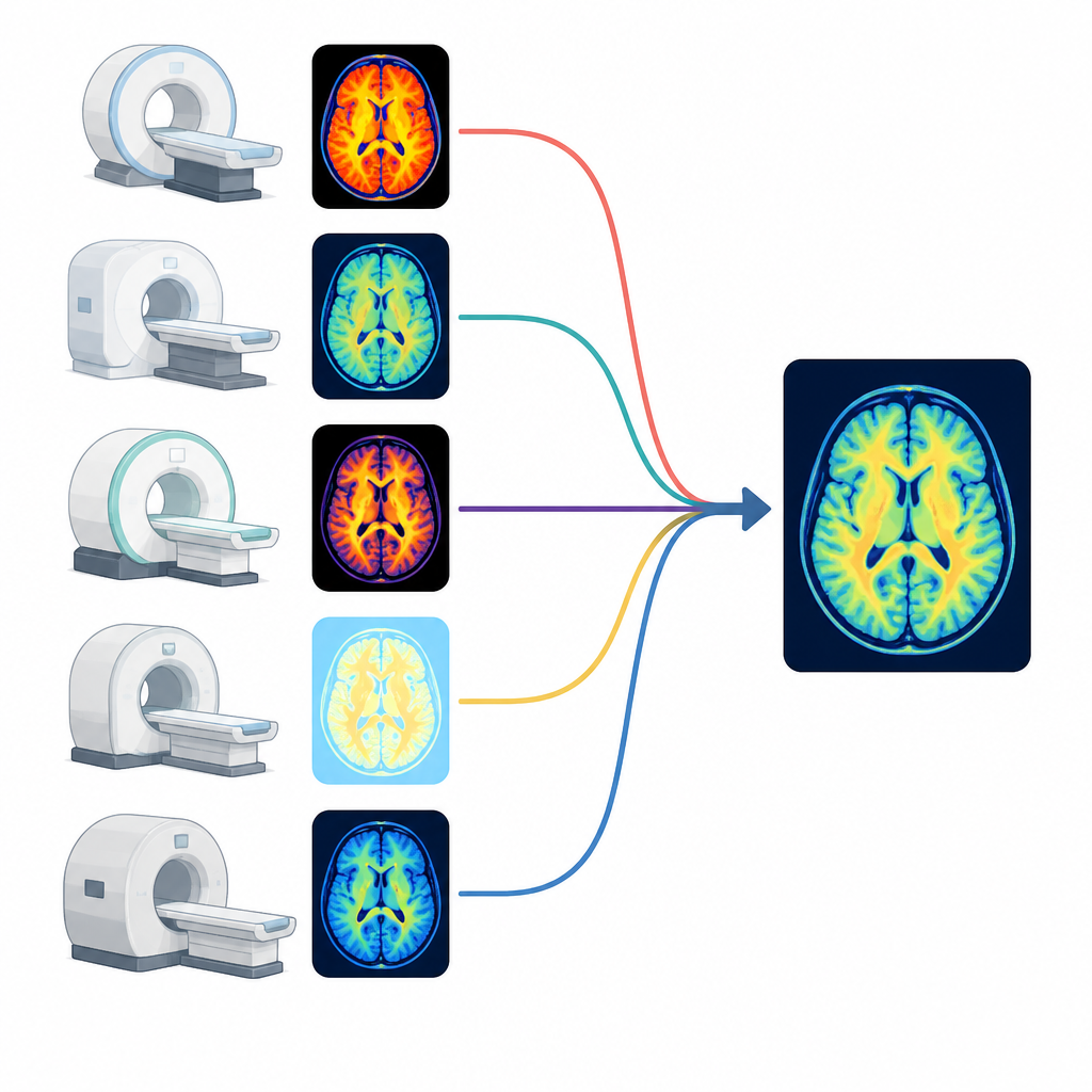
The method was then challenged with tracers and scanners it had never seen. For amyloid imaging with a different tracer, data from two independent centers showed that uncorrected PET‑MRI significantly disagreed with PET‑CT, but the harmonized PET‑MRI became statistically indistinguishable from PET‑CT without retraining. In a larger clinical sample of 420 participants across three sites and four scanner setups, harmonization shrank a key amyloid scale difference between PET‑MRI and PET‑CT from more than 20 units to just over 4. It also brought tau and dopamine transporter measurements into close alignment across platforms. Diagnostic thresholds derived from PET‑CT could be applied to harmonized PET‑MRI with only small adjustments, and no patients near the cutoff were misclassified after correction.
What this means for patients and trials
By aligning PET‑MRI with PET‑CT across different tracers and manufacturers, this deep learning framework turns PET‑MRI from a device that needs its own rules for every tracer into one that can share numbers and cutoffs with PET‑CT. That means patients can benefit from lower radiation while still having results that doctors can compare over years and across centers, including in treatment trials for new Alzheimer’s drugs. Although some tiny systematic differences remain, they are small enough that they do not appear to affect real‑world decisions, making this approach a practical path toward more consistent, safer brain imaging in routine care.
Citation: Wang, J., Zhong, A., Xu, Q. et al. A unified deep learning framework for cross-platform harmonization of multi-tracer PET quantification in neurodegenerative disease. npj Digit. Med. 9, 396 (2026). https://doi.org/10.1038/s41746-026-02570-0
Keywords: PET-MRI harmonization, PET-CT, deep learning, Alzheimer imaging, neurodegenerative disease