Clear Sky Science · en
Real-world unified denoising for multi-organ fast MRI: a large-scale prospective validation
Sharper Scans in Less Time
Magnetic resonance imaging, or MRI, gives doctors detailed views inside the body, but the price is time: patients often need to lie still in a narrow tube for many minutes while the scanner collects enough data to make clear pictures. This study shows how an artificial intelligence system can clean up noisy, quickly acquired MRI images so that scan times drop to around a minute in many cases, without sacrificing the clarity doctors need to make diagnoses.
Why MRI Is Slow and Noisy
MRI machines build images by gathering repeated measurements of signals from the body. Collecting fewer measurements makes the scan faster but also makes the pictures grainy and speckled, which can hide small injuries or subtle brain changes. Existing computer methods can reduce this noise, but they often rely on artificial noise patterns or simplified math that do not truly match what happens in real hospital scanners. Other modern image generators can invent missing details altogether, raising safety concerns when doctors must trust every visible structure.
A Unified Cleaner for Many Body Parts
The researchers tackled this problem by assembling a very large collection of real clinical MRI data. They prospectively gathered 148,930 paired images from six organs, including the head, knee, spine, and shoulder, in six hospitals and across four major MRI brands. For each case there was a noisy fast-scan image and a cleaner reference image produced by slower, more thorough scanning. Using this diverse material, they trained a single denoising system that works directly on standard hospital image files and is designed to plug into existing scanner workflows. 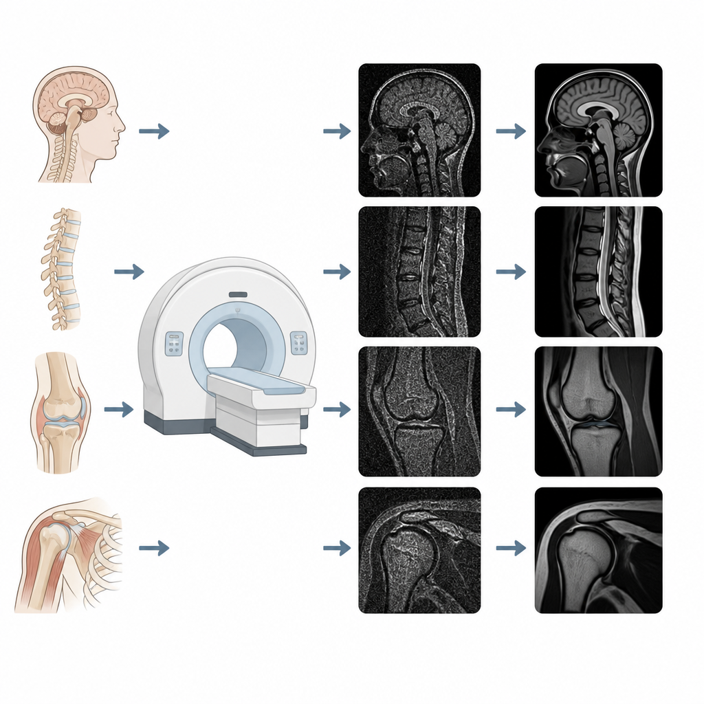
How the Smart Denoiser Works
The heart of the system is a two-part model. One part learns how real scanners and fast protocols degrade images in complex, non-linear ways, rather than assuming simple, uniform noise. The other part is a modern generative model that gradually transforms a noisy picture into a cleaner one. Text information about which organ is being scanned, which type of MRI sequence is used, which manufacturer made the scanner, and how much the scan was sped up is converted into a numerical description that guides the denoising process. During use, the generative model is repeatedly nudged by the learned scanner model so that the final image stays faithful to the original measurements and avoids making up new anatomy. 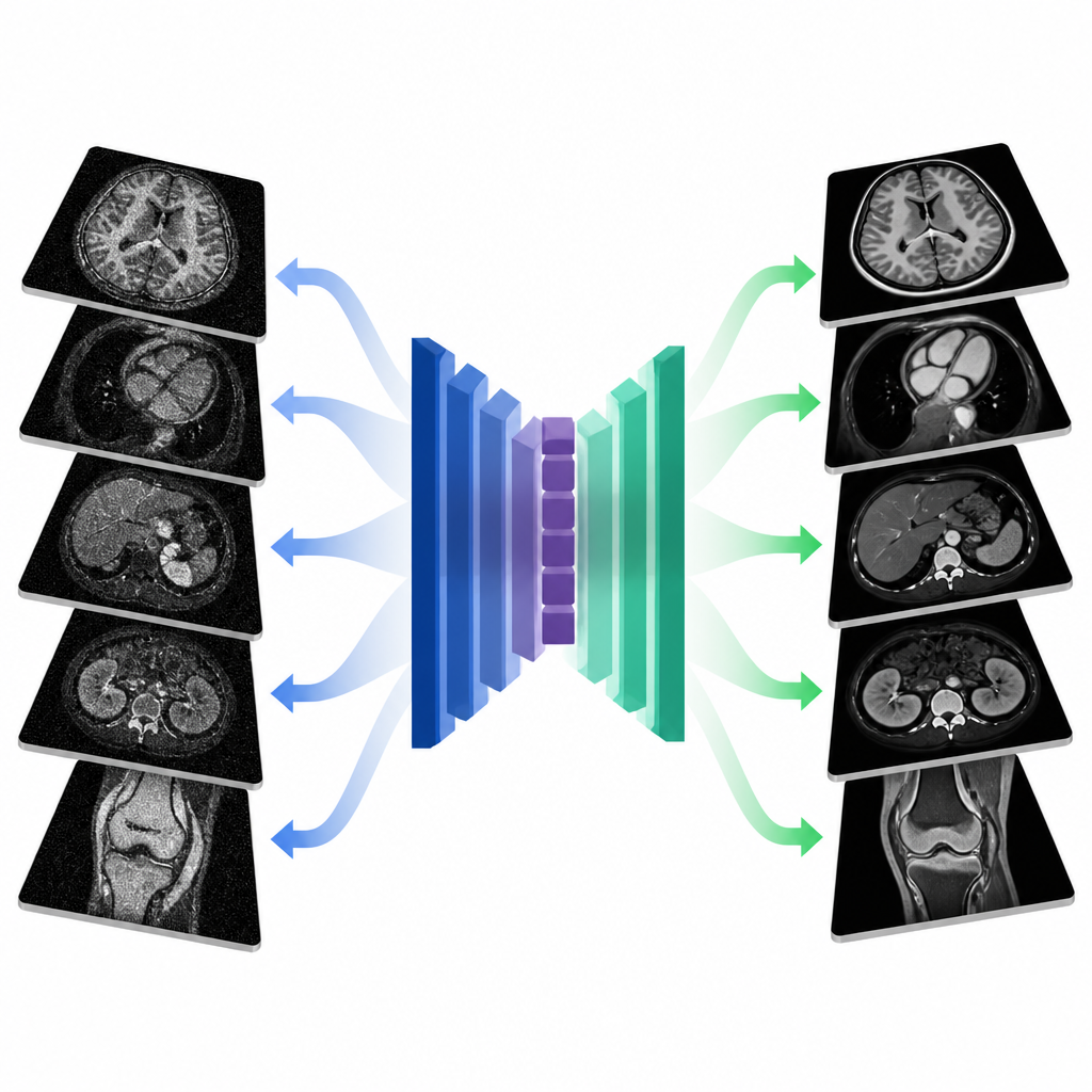
Tests Across Hospitals and Tasks
To judge performance, the team compared their method with five leading denoising approaches on more than twenty thousand test images from their internal cohort. They measured how closely the cleaned images matched the slower reference scans and how well automatic software could segment tissues such as brain gray and white matter or spinal structures. Their system produced sharper, more natural-looking results and improved segmentation accuracy by about seven percentage points compared with using the noisy images alone. They then applied the same trained model, without adjustment, to almost fifty thousand additional scans from four other centers and four scanner vendors. Again, the method produced clearer images and better alignment with reference scans, showing strong generalizability.
What Radiologists Saw
Ultimately, the value of any imaging tool rests on clinical judgement. Two radiologists, blinded to how images were acquired or processed, rated image quality and diagnostic confidence for head, spine, and joint scans. They consistently preferred the denoised images over the noisy fast scans, and their ratings were similar to those for the slower reference images. In focused tests, such as grading small vessel disease in the brain, assessing disc degeneration in the spine, and detecting shoulder or knee injuries, the denoised images matched the reference scans in diagnostic accuracy. Agreements between readers ranged from moderate to excellent, suggesting that the cleaned images support reliable interpretations.
What This Means for Patients
The study concludes that a single AI system can safely clean up fast MRI images from many organs, scanners, and imaging protocols while preserving the essential details that doctors rely on. By allowing clinics to skip repeated signal averaging and still obtain clear pictures, this approach can cut scan times by roughly 30 percent on average, with many routine exams finished in under a minute. For patients, that could mean shorter, more comfortable visits and quicker access to imaging, without giving up the diagnostic confidence of today’s longer MRI scans.
Citation: Shao, Y., Huang, H., Zhang, L. et al. Real-world unified denoising for multi-organ fast MRI: a large-scale prospective validation. npj Digit. Med. 9, 366 (2026). https://doi.org/10.1038/s41746-026-02548-y
Keywords: MRI denoising, fast MRI, medical imaging AI, image quality, diagnostic radiology