Clear Sky Science · en
Advancing diagnostic equity through artificial intelligence chest radiograph screening for osteoporosis in Asian populations
Hidden risk in everyday health checks
Many adults, especially in Asia, are walking around with quietly thinning bones that raise their risk of fractures later in life. Yet most will never receive a bone scan, either because current guidelines focus on older women or because specialized tests are hard to access. This study asks a simple question with big consequences: can a routine chest X-ray, already taken for general health checks, be used with artificial intelligence to flag people whose bones may be dangerously weak, long before a break occurs? 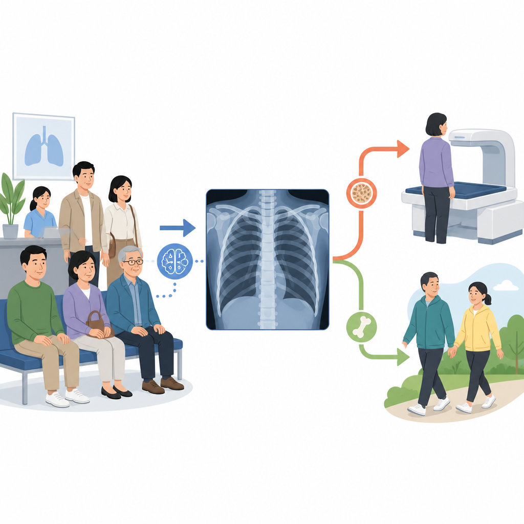
Why weak bones are often missed
Osteoporosis makes bones fragile and prone to fracture, affecting hundreds of millions of people worldwide. In Asia, hip fractures are expected to soar as populations age, yet national screening rules usually target only older women and leave many men and younger adults untested. The standard test, a DXA bone scan, is underused even where it is available, and more than half of serious fractures occur in people whose bone density is not low enough to trigger today’s cutoffs. Body weight is not a reliable shield either: in this study, many people with normal weight still had worrisome bone loss, exposing a blind spot in current screening approaches.
Turning a chest X-ray into a bone check
The research team in Taiwan evaluated a deep learning tool that estimates bone strength from ordinary chest radiographs. These X-rays, taken from the front of the chest, routinely show parts of the spine. The software analyzes this image, predicts bone mineral density, and flags suspected abnormal results for follow-up. To put the tool to the test, the authors used data from 2384 adults who voluntarily attended a preventive health center and had both a chest X-ray and a lumbar spine DXA scan within six months. The group was mostly in midlife, with an average age of about 44 years and a wide range of body sizes typical of East Asian populations.
How well the AI spotted weak bones
When the AI model indicated possible problems, about one in three of those people truly had abnormal bone density on DXA, and it correctly flagged 94 of 118 confirmed cases. Across the whole group, its ability to separate normal from abnormal bone status was very strong, with a high measure of accuracy known as the area under the curve. The tool worked consistently across men and women, younger and older adults, and three body mass index ranges. It was especially good at ruling out disease: if the AI found no problem, the chance of truly normal bones was extremely high. Many of the confirmed cases were women over 50 with normal body weight, a group that might otherwise be overlooked because they do not appear obviously frail. 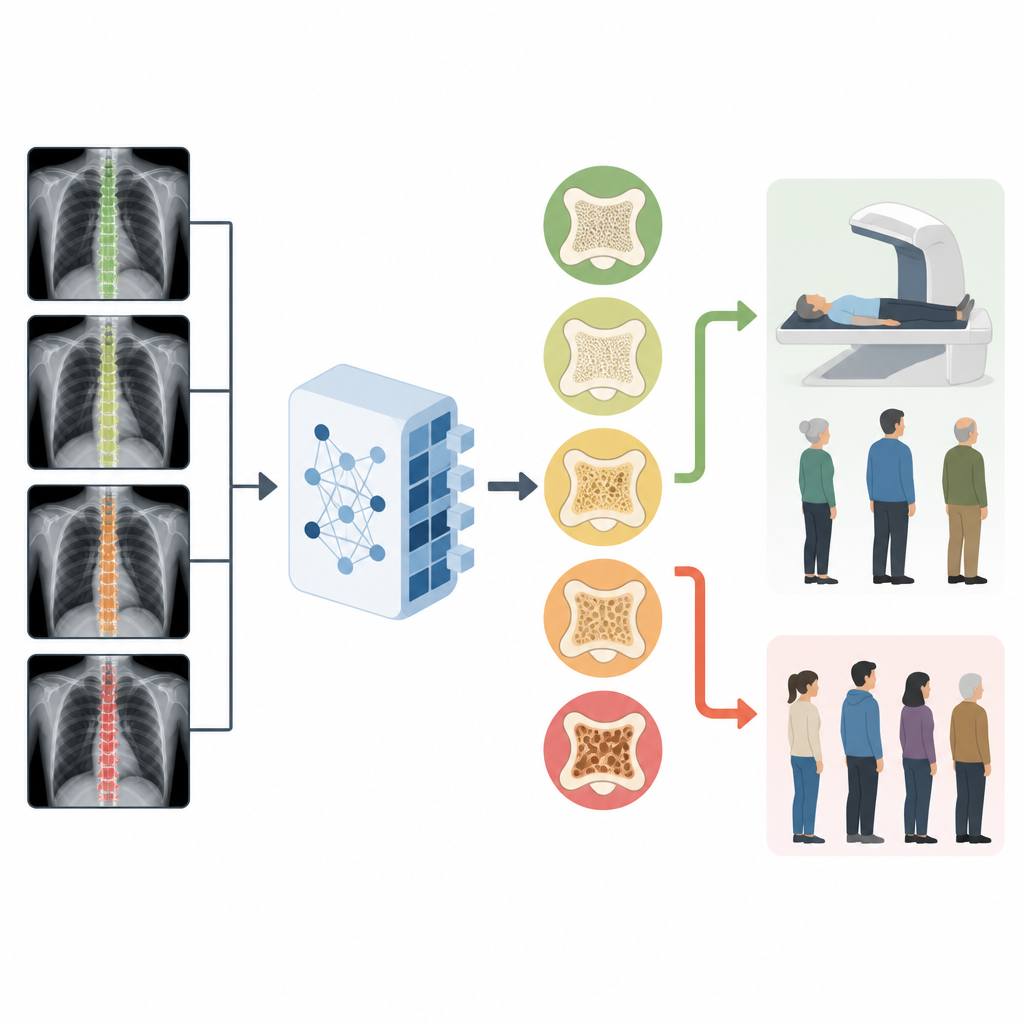
What this could mean for clinics
Because chest X-rays are already performed millions of times each year, adding an automated bone check would not require extra appointments or radiation exposure. Decision analyses in the study showed that using the AI to decide who should get a DXA scan would provide more benefit than sending everyone, or no one, for further testing, particularly among women with normal or low body weight. In practical terms, the software acts as a triage tool: it helps select which patients should be referred for a full bone scan, making better use of limited equipment and clinician attention while reducing missed cases.
Careful steps toward fair and broad use
The authors stress that their findings come from a single health system with relatively few confirmed abnormal cases, so results for some smaller subgroups are uncertain. The tool is not meant to replace DXA or clinical judgment, but to complement them, and it needs further testing in different hospitals, countries, and ethnic groups. Still, the work demonstrates how carefully designed AI can support more equal access to diagnosis by not relying solely on age, sex, or weight. For patients, the message is clear: a familiar chest X-ray could one day double as an early warning system for fragile bones, helping doctors protect more people from painful and life-altering fractures.
Citation: Chen, SH., Chang, RE., Lien, CE. et al. Advancing diagnostic equity through artificial intelligence chest radiograph screening for osteoporosis in Asian populations. npj Digit. Med. 9, 359 (2026). https://doi.org/10.1038/s41746-026-02484-x
Keywords: osteoporosis, bone density, artificial intelligence, chest X-ray, screening