Clear Sky Science · en
Deep learning for fast screening and localization of spinal dural arteriovenous fistulas to enhance clinical workflow
Why this matters for patients and doctors
Back and leg weakness, trouble walking, or bladder problems are often blamed on aging or common spine issues. Yet in some people, the real culprit is a tiny abnormal blood vessel on the covering of the spinal cord—a spinal dural arteriovenous fistula (SDAVF). These fistulas are hard to spot and easy to miss, which can delay treatment and cause permanent nerve damage. This study introduces an artificial intelligence (AI) system that reads spine blood-vessel scans quickly and accurately, helping doctors find these hidden problems with far less effort.
A hidden cause of serious spinal problems
SDAVFs are the most common type of spinal blood-vessel malformation, but they are still rare overall and often overlooked. Their symptoms—gradual leg weakness, numbness, or problems with bladder and bowel control—can mimic many other spinal conditions. If the fistula is not treated in time, high-pressure blood flow can damage the veins around the spinal cord and lead to irreversible disability. Doctors currently rely on an invasive test called digital subtraction angiography, which uses long procedures, radiation, and contrast dye to map the spinal blood vessels in detail. A less invasive scan, CT angiography (CTA), is already used to guide this work, but turning the raw data into usable 3D images is slow, tedious, and highly dependent on the skill and patience of the technician.
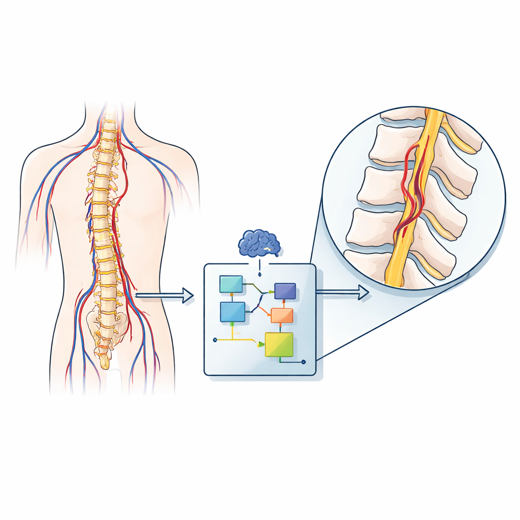
Turning complex scans into clear answers
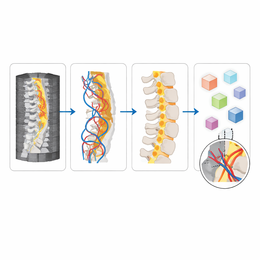
To tackle this bottleneck, the researchers built an automated system called SDAVFdoc that reads whole-spine CTA scans. Instead of asking a human to scroll through more than a thousand thin image slices and manually rebuild the blood vessels, the system breaks the task into several logical steps. First, one deep-learning model outlines the spinal cord itself, shrinking the search area. A second model then searches that region for clusters of abnormal draining veins, the key visual sign of SDAVF. If that cluster reaches a certain size, the system flags the scan as likely positive.
Finding the exact trouble spot
After deciding whether a fistula is likely present, SDAVFdoc still needs to tell the doctor where to look. For this, additional AI models identify the centers of the vertebral bodies from the first thoracic to the fifth lumbar level, then locate the left and right openings where the spinal nerves exit, called the intervertebral foramina. Using this built-in map of the spine, the system narrows down the vertical segment where the draining veins appear, then crops small three-dimensional blocks around each foramen. A final deep-learning model examines these tiny regions and chooses the one most likely to contain the fistula, effectively pointing to a specific side and level of the spine.
Accuracy, speed, and real-world testing
The team trained and tested SDAVFdoc on CTA scans from 718 patients across three hospitals, including both people with confirmed SDAVF and those without. The vein-cluster detection model correctly distinguished SDAVF from non-SDAVF cases with high accuracy, reaching F1-scores above 0.93 in multiple test sets. The final fistula-spotting model also performed strongly, with its ability to separate true from false cases (AUC) consistently around 0.93–0.95. Just as important, two experienced radiologists rated over 90% of the automatically generated vein images as good enough or excellent for diagnosis. In a prospective trial that mirrored daily clinical practice, the AI system cut the average post-processing time per case from about 41 minutes to just over 1 minute and slashed the number of mouse clicks from more than 750 to fewer than 10.
What this means for future care
For patients, the study suggests a future in which dangerous but treatable spinal vessel problems are found more reliably and earlier, before they cause lasting damage. For doctors and technicians, SDAVFdoc offers a way to turn a laborious, expertise-heavy task into an automated background process that delivers clear candidate locations for a fistula. While the system still needs testing on other, rarer spinal vessel diseases and in regions outside the lower spine, it already shows that AI can safely shoulder much of the burden of complex image reconstruction. In practical terms, this tool could help hospitals shorten workups, reduce radiation-heavy procedures, and guide specialists more quickly to the tiny defect that makes the difference between paralysis and recovery.
Citation: Zheng, F., Cao, X., Xu, J. et al. Deep learning for fast screening and localization of spinal dural arteriovenous fistulas to enhance clinical workflow. npj Digit. Med. 9, 296 (2026). https://doi.org/10.1038/s41746-026-02474-z
Keywords: spinal dural arteriovenous fistula, CT angiography, deep learning, medical imaging AI, spinal vascular malformations