Clear Sky Science · en
Automated interpretation of fetal cardiac function evaluation from the echocardiogram
Why this matters for expectant families
Prenatal scans do far more than reveal a baby’s profile—they can hint at how well the tiny heart is working long before birth. But today, measuring fetal heart function from ultrasound videos is slow, demanding work that depends heavily on the skill of a few specialists. This study describes a new artificial intelligence (AI) system that can automatically read these heart scans, promising faster, more consistent assessments that could help doctors spot trouble earlier and follow high‑risk pregnancies more closely.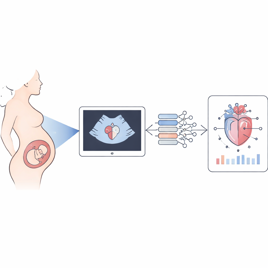
Turning heart scans into numbers
When an expectant parent has a fetal heart ultrasound, the machine records moving images of the beating heart. Experts then pause the video at just the right moments and carefully trace the walls and chambers to calculate how well the heart pumps. The researchers in this study built an AI “workflow” to carry out that entire process automatically. Their system watches the moving ultrasound, finds the four‑chamber view of the heart, outlines key structures such as the upper and lower chambers and the chest, and then converts those outlines into more than 70 different measurements that describe the size, shape, and pumping strength of the fetal heart.
Training the system on many real pregnancies
To teach the AI what a fetal heart looks like across many situations, the team used over fifty thousand labeled images from nearly two thousand normal fetal heart videos, collected at one major hospital. They then tested it further on additional normal cases from two other hospitals and on 83 fetuses with various heart‑related or growth problems. In all, the dataset covered gestational ages from 18 to almost 38 weeks and a wide variety of fetal positions and scan conditions. Experienced sonographers carefully annotated the images used for training and never took part in the later comparisons, helping to ensure an unbiased evaluation.
Matching and even smoothing out expert opinions
To judge whether the AI could stand in for humans, the researchers compared its measurements with those made independently by two experienced sonographers, both by hand and using a popular semi‑automatic tool called Fetal Heart Quantification. Across right‑sided, left‑sided, and whole‑heart measurements, the AI’s results agreed with each expert better than the experts agreed with one another. Differences between AI and human readings were smaller than typical differences between two human readers, and statistical tests indicated that the AI’s variability was actually lower. Importantly, this held true not only in routine pregnancies but also in scans from fetuses with heart or growth abnormalities, and on data from outside hospitals, suggesting that the system can generalize beyond its original training site.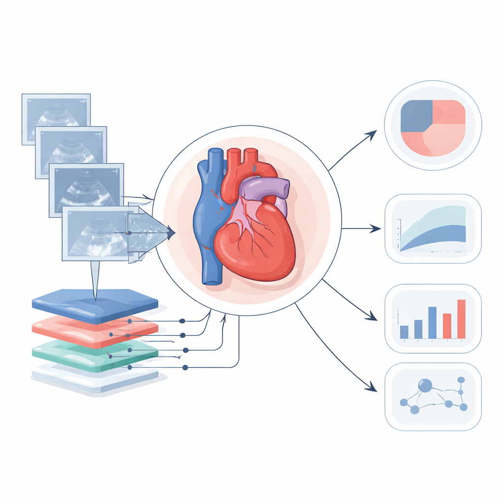
Linking heart function to overall fetal growth
Beyond one‑off measurements, doctors also need to know whether a given baby’s heart is performing as expected for its stage of growth. The team used the AI’s output from 1,385 normal pregnancies to build a “Z‑score” model—a way of expressing how far a particular measurement sits above or below the typical value after accounting for growth. They tested 44 different equations that linked heart measurements to commonly collected fetal size indicators such as head and abdominal circumference, femur length, and estimated weight. Estimated fetal weight turned out to best capture how many structural heart measurements change as babies grow. Most normal fetuses fell comfortably within the predicted ranges, while several fetuses with serious conditions, including narrowed aortas and growth restriction, showed heart measurements that clearly fell outside the expected band.
What this means for prenatal care
In plain terms, this study shows that a computer system can watch a standard fetal heart ultrasound, automatically trace the chambers, and produce a detailed report that matches or even improves upon the consistency of human experts. By wrapping these measurements into growth‑adjusted scores, the tool can help highlight fetuses whose hearts may be under unusual strain or developing atypically. While more work is needed to test the system in additional hospitals, on more types of ultrasound machines, and in more complex heart defects, the findings point toward a future in which detailed fetal heart function can be measured quickly, objectively, and routinely during pregnancy, rather than only when a specialist has ample time.
Citation: Huang, C., Zhang, L., Xie, B. et al. Automated interpretation of fetal cardiac function evaluation from the echocardiogram. npj Digit. Med. 9, 334 (2026). https://doi.org/10.1038/s41746-026-02381-3
Keywords: fetal echocardiography, artificial intelligence, prenatal ultrasound, cardiac function, Z-score model