Clear Sky Science · en
Construction of virtual whole eye model based on ultra-widefield optical coherence tomography in myopia
Why the shape of the eye matters
Blurry distance vision, or myopia, is rapidly becoming one of the world’s most common vision problems, especially among children and young adults. In severe cases, it can lead to eye diseases that threaten sight for life. This study introduces a new way to build a full three-dimensional “virtual eye” using a quick, clinic-friendly scan instead of an expensive MRI machine. By turning routine images into detailed digital eye models, doctors could one day track how myopia reshapes the eye over time and tailor treatments before lasting damage occurs.
From hospital scanner to virtual eye
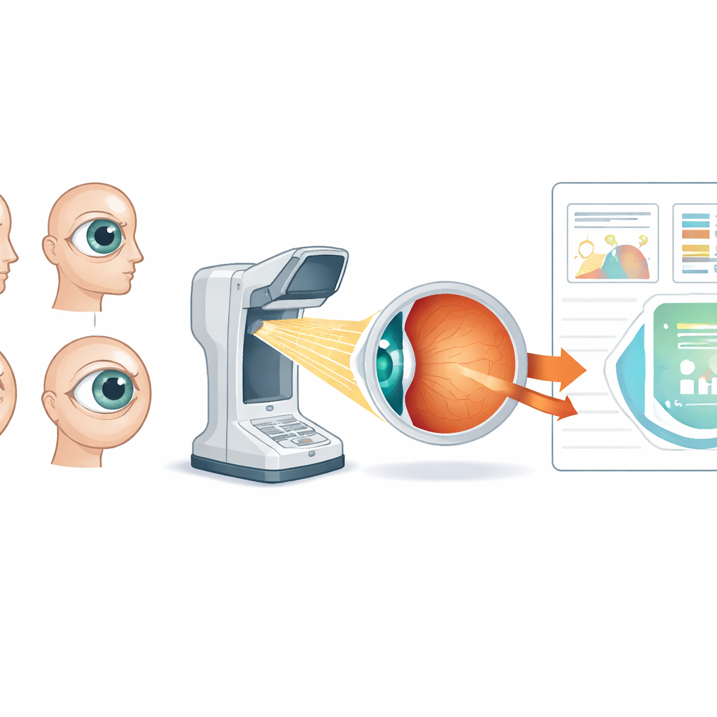
The researchers set out to solve a practical problem: MRI can capture the entire eyeball and show its true shape, but it is costly, time-consuming, and not suitable for large numbers of patients or repeated checkups. In contrast, a newer imaging method called swept-source optical coherence tomography (SS-OCT) is already common in eye clinics and can scan a very wide area of the retina in just a few seconds. The team designed a special “radial” scanning pattern and custom software, called CET-1, that stitches SS-OCT images from the front and back of the eye into a single whole-eye model, including the cornea, lens, and retina.
Checking the new model against MRI
To see whether this virtual eye was trustworthy, the scientists compared CET-1 models with MRI-based models in 70 eyes from adults whose vision ranged from normal to extreme myopia. They aligned both models in three dimensions using landmarks like the cornea, the center of sight (the fovea), and the optic nerve head. Then they measured how far each point on the CET-1 surface was from the corresponding MRI surface. Across a wide span of eye lengths, the average difference was less than half a millimeter and did not grow as eyes became more elongated. In the most important central region, which includes the macula and optic nerve, the match was even tighter, while larger mismatches were mostly confined to the far periphery where current scans are less complete.
How myopia reshapes the back of the eye
With a reliable virtual model in hand, the team examined how the eye surface bends and bulges as myopia worsens. Using a mathematical measure called Gaussian curvature, they mapped how steeply the retina curves at each point. The CET-1 models revealed that with increasing myopia, the back of the eye becomes progressively more curved and irregular, especially in highly and extremely myopic eyes. These patterns were visible in eyes with severe outpouchings of the wall, known as posterior staphylomas, and the curvature values differed clearly from those in eyes with normal vision. Such curvature maps may offer a new way to flag eyes at high risk for serious complications like macular damage or retinal splitting.
Front-of-the-eye changes in growing myopia
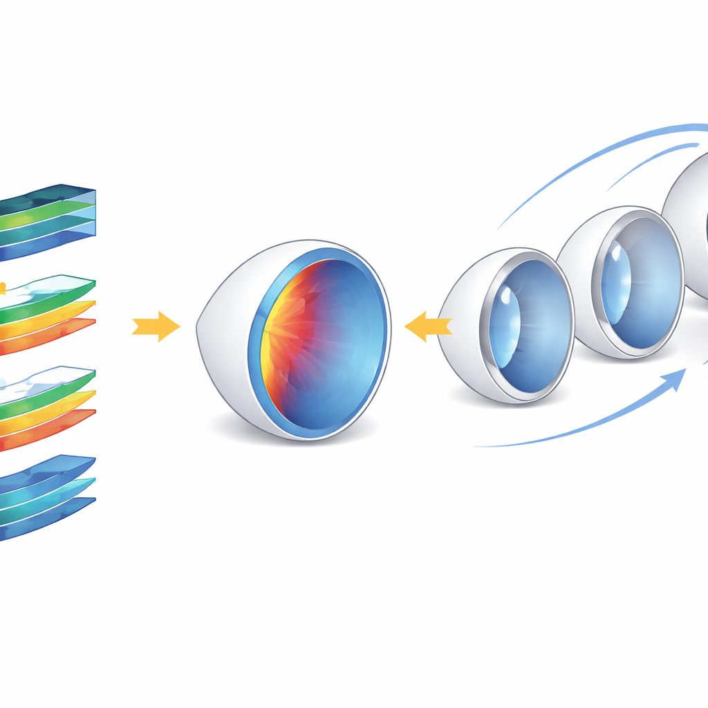
The virtual models also captured subtle changes in the eye’s front structures, which standard retinal studies often ignore. As myopia severity increased, several measures of the anterior chamber—the space between cornea and lens—grew larger, including its depth, volume, and the angle where the cornea meets the iris. At the same time, the “lens vault,” or how far the lens projects forward, tended to shrink in more myopic eyes. These shifts may help explain why people with high myopia are more prone to other eye diseases, such as certain types of glaucoma and cataracts, by altering how fluid drains from the eye or how the lens ages and loses focusing power.
Toward digital twins for personalized eye care
By showing that a fast, clinic-ready scan can recreate the overall eyeball shape almost as accurately as MRI, this work paves the way for building “digital twins” of patients’ eyes: living virtual models that can be updated at each visit. Such twins could help researchers study how myopia develops, test how different treatments might change the eye’s shape, and identify early warning signs of dangerous complications long before vision is lost. While the current study focuses on adults and still relies on some estimated regions, it demonstrates a powerful new tool for large-scale, personalized monitoring of myopia in the real world.
Citation: Tang, X., Luo, N., Chen, C. et al. Construction of virtual whole eye model based on ultra-widefield optical coherence tomography in myopia. npj Digit. Med. 9, 298 (2026). https://doi.org/10.1038/s41746-026-02376-0
Keywords: myopia, virtual eye model, optical coherence tomography, digital twin medicine, ocular morphology