Clear Sky Science · en
A comprehensive foundation model for generalizable cytogenetics in precision oncology with CHROMA
Why looking at chromosomes still matters
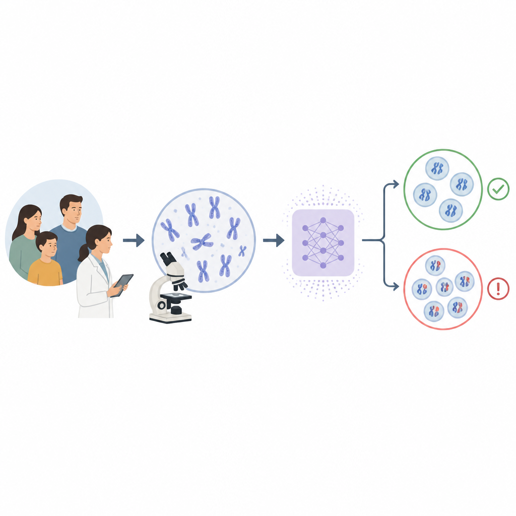
When doctors investigate genetic diseases and many cancers, they still rely on pictures of a patient’s chromosomes to spot missing, extra, or rearranged pieces of DNA. This careful visual checking is slow, depends on rare specialists, and can be especially hard in hospitals with limited staff or older equipment. The study introduces CHROMA, an artificial intelligence system designed to help read these chromosome images more quickly and reliably, aiming to make advanced genetic testing easier to access around the world.
A new digital helper for chromosome reading
CHROMA is presented as a foundation model, a versatile AI system trained on a very large and varied collection of data so it can handle many related tasks. In this case, the team fed CHROMA more than four million chromosome images from over 84,000 patients. These images show the familiar X shaped structures arranged during cell division, and they include both normal patterns and a wide range of genetic problems. Instead of focusing on one narrow job, CHROMA is built to recognize many kinds of chromosome changes in a single framework, from extra or missing copies to broken and rejoined fragments, including rare forms that are seldom seen in routine practice.
Training an AI to see what experts see
To teach CHROMA without demanding line by line human labels for every image, the researchers used self supervised learning. They asked the model to fill in missing parts of chromosome pictures and to clean up noise, which forces it to learn the fine detail of banding patterns and shapes that human experts rely on. Later, they added a more modest amount of carefully labeled data so CHROMA could link these visual cues to specific types of abnormalities. Across several test sets, the system correctly told apart the 24 different human chromosomes with very high accuracy and outperformed existing computer methods, even when images were blurry, stained unevenly, or crowded with overlapping chromosomes.
Finding rare and subtle chromosome changes
Beyond simple counting of chromosomes, many diseases involve more subtle rearrangements, such as pieces swapping places, flipping, or forming rings. These problems are harder to spot and may be very rare, which makes them difficult to capture in training data. The team assembled special datasets that included both common changes and a small number of unusual cases. CHROMA was able to detect abnormal cells reliably across all of these groups and showed much less drop in performance when data were scarce compared with other AI approaches. The model also naturally grouped different types of abnormalities into distinct clusters inside its internal representation, suggesting it has learned a rich picture of how chromosomes can go wrong, even when it is only asked to separate normal from abnormal.
Building safety into AI supported diagnosis
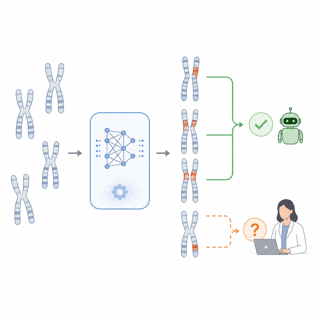
Because mistakes in genetic diagnosis have serious consequences, the researchers added a risk control strategy to CHROMA. Instead of forcing a confident answer on every cell, the system estimates how sure it is about each prediction. If a case looks clear, CHROMA provides a normal or abnormal decision. If the pattern is unusual or the model is uncertain, it automatically flags the cell for a human specialist to review, especially for very rare patterns like chromosomes with three attachment points. In testing, this approach nearly eliminated missed abnormal cells by steering doubtful cases to experts, acting more like a careful triage assistant than an automatic replacement for human judgment.
What this means for patients and clinics
In simple terms, the study shows that a carefully trained AI can scan huge numbers of chromosome images, find a wide range of genetic problems, and know when to ask for help. CHROMA reduces the amount of detailed labeling needed from experts, handles noisy images better than earlier tools, and embeds safeguards to avoid silent errors. While it does not take over the specialist’s role in fully describing each abnormality, it can speed up screening and free experts to focus on the hardest cases. If validated in real time hospital workflows, such systems could bring more accurate chromosome based testing to cancer clinics and genetics services, including those in regions where trained cytogeneticists are in short supply.
Citation: Yang, C., Dai, W., Zhang, Y. et al. A comprehensive foundation model for generalizable cytogenetics in precision oncology with CHROMA. npj Precis. Onc. 10, 187 (2026). https://doi.org/10.1038/s41698-026-01383-4
Keywords: chromosome imaging, precision oncology, medical AI, genetic abnormalities, cytogenetics