Clear Sky Science · en
Distinct metabolic profiles in lung adenocarcinomas presenting as solid or ground-glass opacities
Why lung scan spots are not all the same
When doctors perform CT scans of the lungs, they often find small spots that can signal early lung cancer. Some appear hazy, like frosted glass, while others look dense and solid. Clinically, these different appearances predict how dangerous a tumor is—but why they behave so differently has been a mystery. This study digs into the chemistry inside these tumors, revealing how their energy and fat-processing pathways may explain why some grow slowly and others act more aggressively. 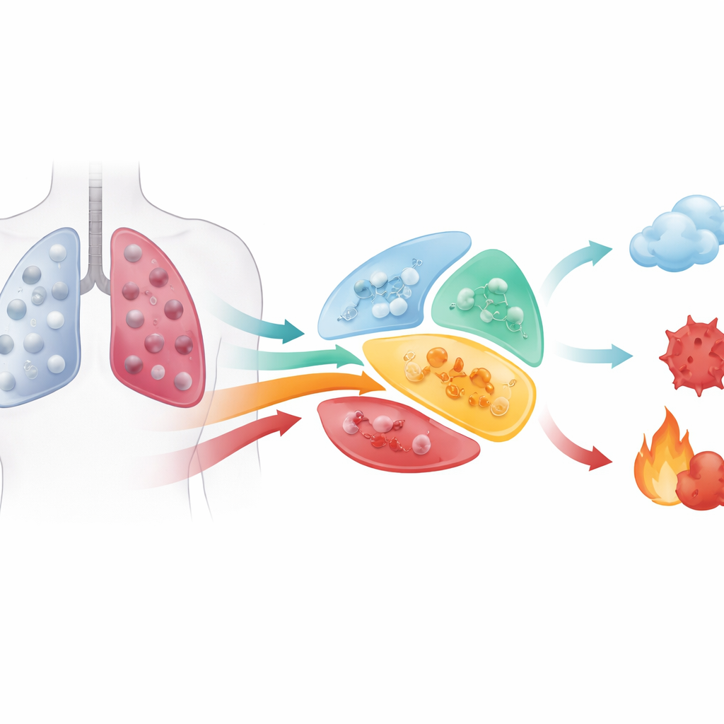
Different kinds of lung spots, different futures
Lung adenocarcinoma is the most common form of lung cancer. On scans, early tumors usually fall into three categories: pure ground-glass opacities (GGOs), which are hazy; solid nodules (SNs), which are dense; and mixed ground-glass opacities (mGGOs), which combine both features. Patients with pure GGOs tend to do remarkably well after surgery, while those with solid nodules face a higher risk of recurrence and spread. Mixed nodules fall somewhere in between. These patterns have shifted how doctors treat early lung cancer, but until now, most explanations focused on genes and immune cells rather than the actual chemical fuel and building blocks inside the tumors.
Mapping the chemistry of early lung cancers
The researchers analyzed 262 tissue samples from 165 patients with early- to mid-stage lung adenocarcinoma. They measured hundreds of small molecules (metabolites) using high-resolution mass spectrometry, examined gene activity with RNA sequencing, and profiled bacteria living within tumors using 16S rRNA sequencing. By linking genes to enzymes, reactions, and metabolites, they built a “wiring diagram” of the tumor’s chemical networks. Overall, tumor tissue looked very different from nearby normal lung, especially in pathways that handle fats and related molecules called glycerophospholipids, key components of cell membranes. This confirmed that reprogrammed metabolism is a central feature of these cancers.
How hazy and solid tumors diverge
Among many clinical factors—age, sex, smoking history, tumor size, and imaging markers—the single strongest divider of metabolic patterns was whether a tumor appeared as a pure GGO or a solid nodule. Tumors with similar density clustered together in chemical space, even when they arose in the same patient. Surprisingly, in mixed nodules, the ground-glass and solid parts looked metabolically alike and closely resembled pure GGOs, not solid nodules. When the authors examined which pathways differed most between pure GGOs and solid nodules, one stood out: linoleic acid metabolism, a branch of fat processing that shapes cell membranes and produces signaling molecules tied to inflammation and growth.
Membrane fats, key enzymes, and tumor behavior
Zooming in on linoleic acid metabolism, the team found that solid nodules had altered levels of certain phospholipids, particularly a drop in a molecule called phosphatidylcholine (32:0) and a rise in a related compound containing polyunsaturated fatty acids. Genes encoding cytosolic phospholipase A2 (cPLA2)—an enzyme that clips fatty acids from membrane lipids—were more active in solid nodules, hinting that this enzyme helps drive a more aggressive state. To test this idea, the researchers used lung cancer cell lines and patient-derived organoids grown in the lab. Blocking cPLA2 with a chemical inhibitor reduced cell invasion and proliferation, while enriching cell membranes with phosphatidylcholine (32:0) using specially prepared liposomes also slowed growth and spread. These experiments suggest that tweaking membrane composition and its processing enzymes can directly influence how invasive lung cancer cells become. 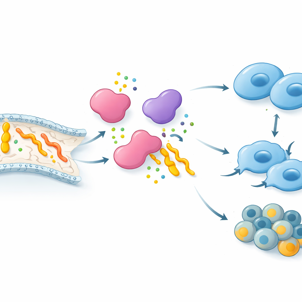
The role of microbes and what comes next
The team also asked whether bacteria living inside tumors help explain the differences between hazy and solid lesions. Although certain bacterial types were more common in one group or the other, the overall community structure and diversity were similar, and microbes did not appear to be the main drivers of the metabolic split between GGOs and solid nodules. Still, some strong links emerged between specific bacteria and individual metabolites, hinting at a more subtle microbe–metabolism interplay that future long-term studies may unravel.
What this means for patients and care
For non-specialists, the major message is that a nodule’s appearance on a CT scan reflects deep chemical differences inside the tumor. Hazy ground-glass lesions tend to have a “gentler” metabolic profile, whereas dense solid nodules show membrane and fat-processing changes that favor invasion and rapid growth. The study points to phospholipids and enzymes like cPLA2 as potential targets for new diagnostics, imaging tracers, and treatments that could be tailored to the tumor’s subtype. In the long term, understanding and manipulating these metabolic signatures may help doctors better predict which early lung cancers need aggressive treatment and which can be managed more conservatively.
Citation: Li, B., Wang, D., Wang, Y. et al. Distinct metabolic profiles in lung adenocarcinomas presenting as solid or ground-glass opacities. npj Precis. Onc. 10, 174 (2026). https://doi.org/10.1038/s41698-026-01378-1
Keywords: lung adenocarcinoma, ground-glass opacity, tumor metabolism, phospholipids, precision oncology