Clear Sky Science · en
Radiation dose has no significant impact on CT-based bone mineral density measurements in a large-animal model
Why this matters for everyday health
As people live longer, weak bones and fractures become a growing concern, yet many at risk never get a dedicated bone test. This study explores whether doctors can safely “recycle” the many CT scans already done for other reasons to estimate bone strength, without worrying about how much radiation was used for each scan.
Bones, scans and a chance to spot hidden risk
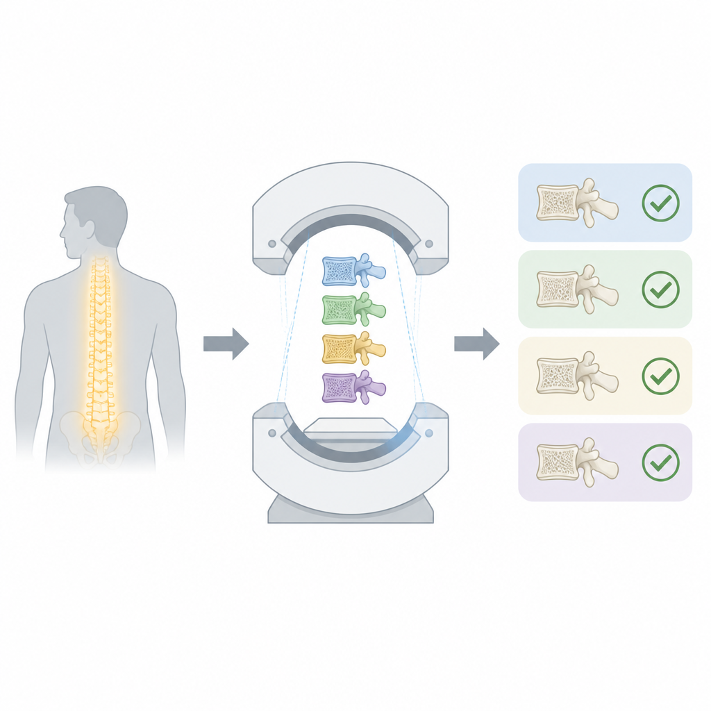
Bone mineral density, a measure of how solid bones are, helps predict frailty, fractures and even survival in serious illnesses. Today, it is usually measured with a special low dose X ray test that gives only a flat, two dimensional view and requires a separate appointment. Modern CT scanners already create detailed three dimensional pictures of the body for many patients. If those same scans could also give reliable information about bone strength, doctors could check for hidden osteoporosis “opportunistically” during routine imaging, saving time, cost and extra radiation.
Testing bone readings at very different radiation levels
One concern is that changing the radiation dose on a CT scanner alters image noise, which might distort bone density readings. To test this, researchers studied twenty minipigs, whose spine structure is similar in size and shape to that of humans in key ways. Each animal underwent several CT sessions, and in every session the same part of the spine was scanned five times in a row using doses ranging from full standard dose down to just five percent of that level. In total, this produced nearly six hundred scans under tightly controlled conditions on the same scanner.
Smart software to read the spine
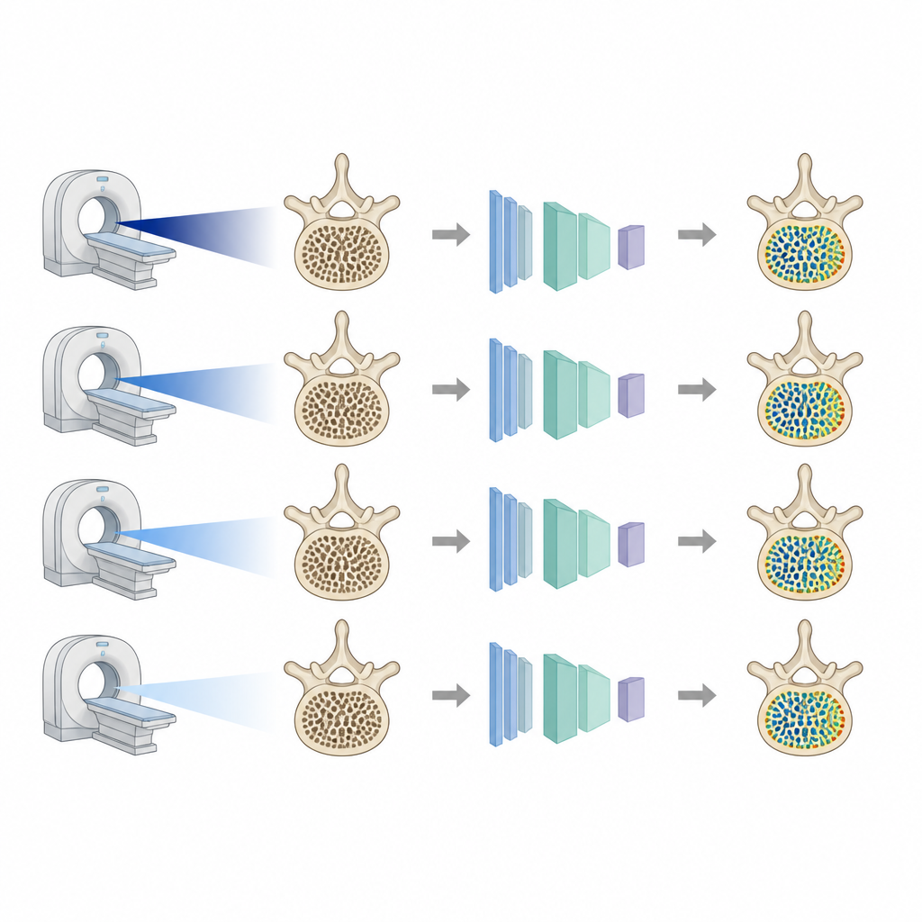
Instead of asking experts to outline each bone by hand, the team used advanced machine learning software trained to recognize body structures in CT images. They adapted an existing human body analysis network to the anatomy of the minipigs by manually marking thousands of slices to teach the system where each vertebra begins and ends. The software then automatically measured the average brightness of the ninth thoracic vertebral body, which reflects bone density, and also placed small regions of interest inside the spongy inner bone while avoiding the hard outer shell. These automatic segmentations matched manual outlines very closely, showing the method was precise.
Bone readings stay steady as dose drops
When the researchers compared bone density measurements across the different dose settings, they found that the values were almost identical. Even at just five percent of the standard dose, the average measurements for the whole vertebra and for the inner spongy bone differed from the full dose scans by well under two percent, and statistical tests found no meaningful differences. As expected, values from the inner bone were slightly lower than those from the entire vertebra, but this gap was consistent across all dose levels, highlighting the need to compare like with like when tracking bone changes over time.
What this means for future checkups
The study shows that in this large animal model, bone density estimates from CT images remain stable even when radiation is sharply reduced. This suggests that, within similar technical limits, doctors may be able to compare scans acquired with different CT dose settings when assessing bone health and spotting osteoporosis during routine imaging. While further work in human patients and with other scanners is needed, the findings support the idea that information already hidden in CT scans could be safely reused to help prevent fractures without extra tests.
Citation: Harmes, J.C., Holtkamp, M., Straus, J. et al. Radiation dose has no significant impact on CT-based bone mineral density measurements in a large-animal model. Sci Rep 16, 16570 (2026). https://doi.org/10.1038/s41598-026-55169-6
Keywords: bone density, CT scans, radiation dose, osteoporosis screening, machine learning