Clear Sky Science · en
Viscoelastic profiling of rare pediatric extracranial tumors using multifrequency MR elastography: a pilot study
Why the softness of tumors matters
When a child is found to have a tumor, doctors want every bit of information they can get without adding extra pain or risk. This study explores a way to “feel” how firm or fluid a tumor is from the inside, using a standard MRI scanner. By gently vibrating the body and watching how those vibrations move through tissue, the researchers tested whether they could learn more about rare tumors outside the brain in children, and whether these measurements relate to how aggressive the tumors are.
A new way to feel tissue from the inside
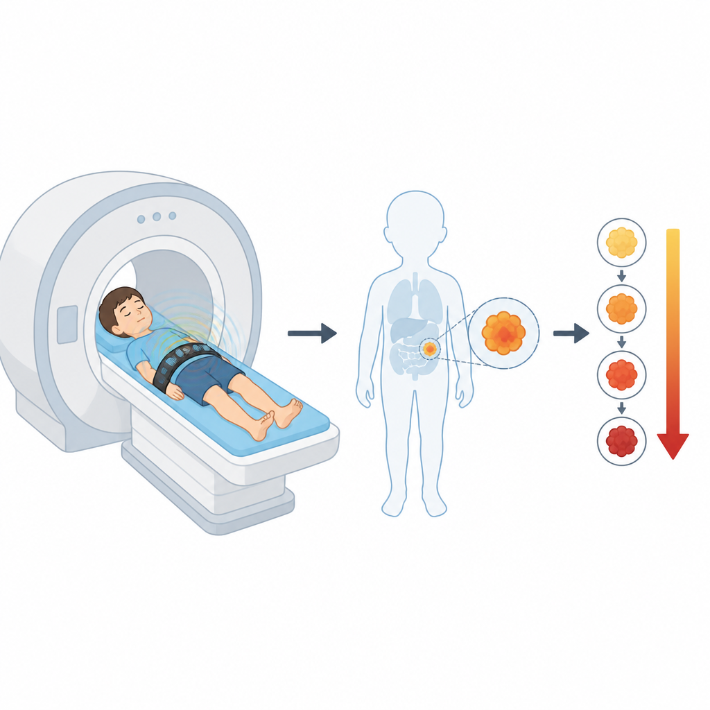
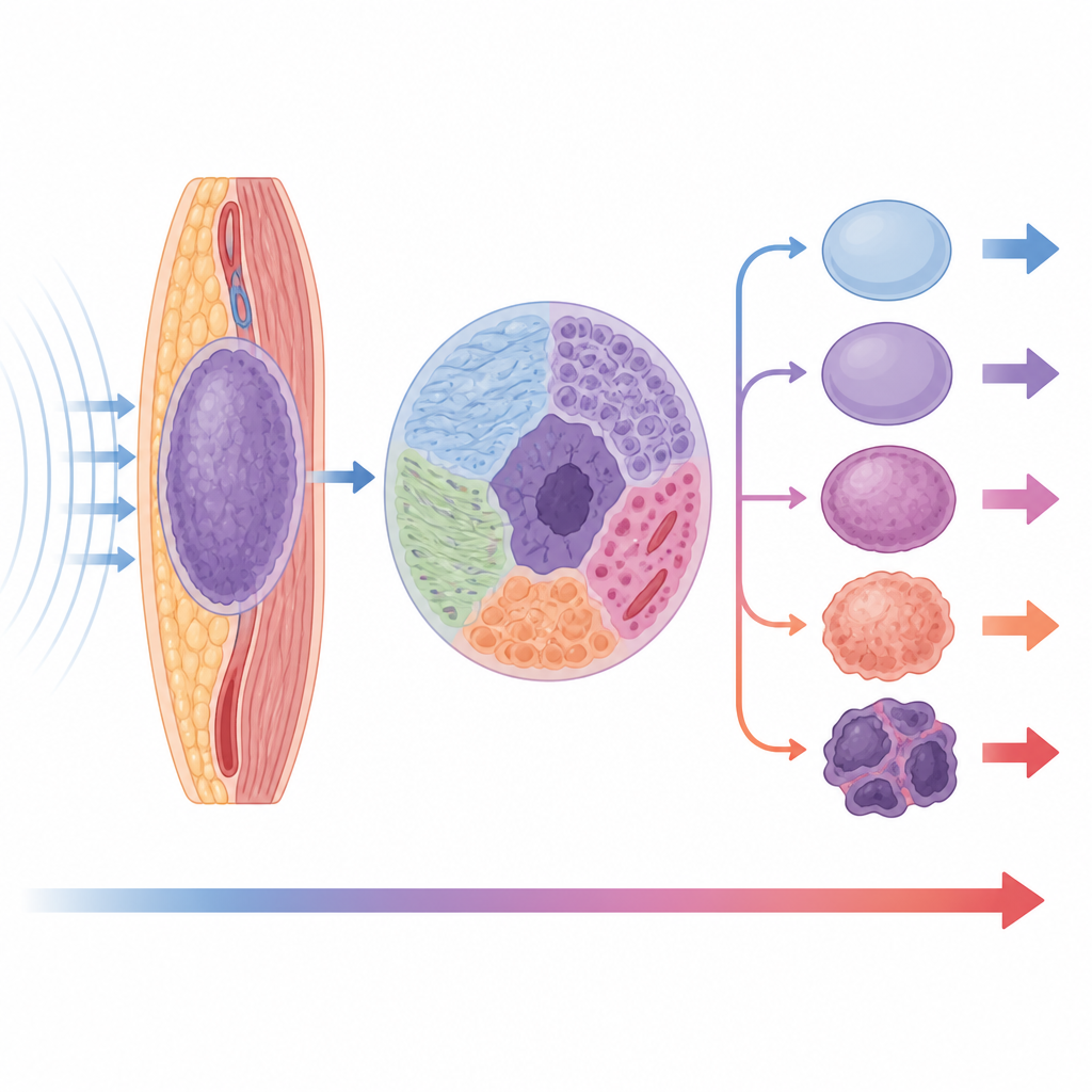
The technique at the heart of this work is called magnetic resonance elastography, or MRE. Instead of relying only on how a tumor looks, MRE adds a mechanical dimension: how stiff it is and how easily it flows or deforms. During an MRI exam, soft air-powered pads placed on the child’s body create gentle waves that travel through organs and tumors. The MRI scanner tracks these waves, and computer algorithms turn the wave patterns into colorful maps that show stiffness and a related property called fluidity, which reflects how viscous or “gooey” the tissue behaves on a microscopic level.
Who was scanned and how
The team studied ten children, from four months to fifteen years old, each with a different solid tumor outside the brain. These included high- and low-risk neuroblastomas in the adrenal glands, sarcomas in bone and soft tissue, a liver tumor, a kidney tumor, a nerve sheath tumor, and a fatty tumor. For each child, MRE was added to a routine MRI scan and took less than five minutes. The vibrations were applied at several low sound-like frequencies, and three-dimensional wave patterns were captured across multiple slices. The researchers also used another common MRI method called diffusion-weighted imaging, which tracks how freely water molecules move within the tumor.
What the waves revealed about tumors
From the wave data, the scientists calculated how fast shear waves moved through each tumor, a measure that reflects stiffness, and how much the waves lagged behind the driving motion, which reflects fluidity. They then grouped the tumors into four risk levels, from benign to high-risk malignant, based on established clinical and biological criteria. In general, tumors in the higher risk groups tended to be stiffer, more fluid-like, and more irregular in their mechanical properties from place to place. Benign tumors such as the lipoma and schwannoma showed the lowest stiffness and fluidity values, while aggressive tumors like rhabdomyosarcoma and high-risk neuroblastoma showed higher values and greater patchiness.
Linking movement of water and mechanical feel
The researchers also compared the mechanical maps from MRE with diffusion measurements, which are already used to help separate benign from malignant tissue. Tumors that were stiffer and more fluid-like generally showed more restricted water movement, a pattern often associated with dense, highly cellular cancers. This relationship was not perfect: cystic or partly broken-down tumors, and those already altered by treatment, could behave differently. Still, the broad trend suggests that combining how tissue resists deformation with how water diffuses through it may provide a richer picture of tumor structure than either method alone.
What this could mean for children with tumors
This pilot study shows that multifrequency MRE can be safely woven into standard pediatric MRI sessions and can produce meaningful maps of how rare tumors feel from the inside. The early results hint that stiffer, more fluid-like, and more mechanically uneven tumors are often those classified in higher clinical risk groups. While the study is small and diverse, and the authors stress that the findings are exploratory, it raises the possibility that future care could use these noninvasive “touch” measurements alongside existing imaging to better characterize tumors and monitor how they respond to treatment, all without additional needles or radiation.
Citation: Metz, C., Veldhoen, S., Deubzer, H.E. et al. Viscoelastic profiling of rare pediatric extracranial tumors using multifrequency MR elastography: a pilot study. Sci Rep 16, 16588 (2026). https://doi.org/10.1038/s41598-026-55127-2
Keywords: magnetic resonance elastography, pediatric tumors, tumor stiffness, diffusion MRI, tumor risk