Clear Sky Science · en
Uncertainty estimation and probabilistic skull shape reconstruction using bayesian neural networks
Why this work matters for patients and doctors
When surgeons repair damaged skulls after injury or disease, they often rely on computer models to fill in missing bone and design custom implants. These digital reconstructions can look very precise, but they usually hide an important fact: there is more than one way to rebuild a missing piece, and the computer’s answer may be less certain in some regions than others. This study explores how to not only reconstruct the skull in three dimensions but also to show, in a transparent way, where the computer is confident and where it is unsure. 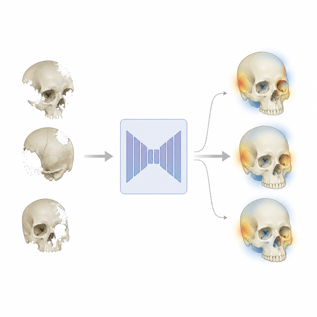
From missing pieces to complete skulls
The authors focus on three related tasks that matter in real clinical scenarios. First, cranial reconstruction aims to restore the upper part of the skull when pieces of bone are missing, for example after surgery that relieves pressure on the brain. Second, facial reconstruction tries to rebuild broken or absent facial bones, a task that is trickier because human faces vary greatly. Third, skull shape super-resolution sharpens coarse, blocky scans into detailed, smooth bone surfaces. In all three cases, the same basic challenge appears: from incomplete or low-quality data, there are several anatomically reasonable ways to fill in the gaps, so a single fixed solution can be misleading.
Teaching a network to admit what it does not know
To tackle this, the researchers adapt a popular image-analysis design called a U-Net, which processes three-dimensional images through a contracting path that captures global structure and an expanding path that recovers detail. In their version, parts of the expanding path are made probabilistic, so the internal settings of the model are treated not as fixed numbers but as values that can vary according to learned probability distributions. During training, the network learns which settings are tightly constrained by the data and which can safely vary. During testing, the same skull can be passed through the model many times, each time sampling slightly different internal settings. By averaging and comparing these repeated reconstructions, the team derives both a best-guess skull and a voxel-wise map of uncertainty. 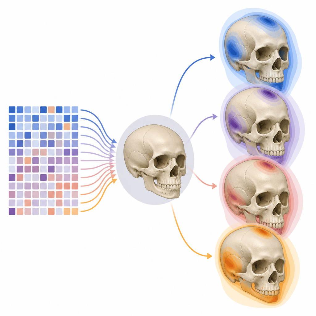
What the model reveals about skull variation
Applying this Bayesian U-Net to a public cranial surgery dataset, the authors show that the model can generate a family of realistic skull reconstructions rather than a single rigid answer. The main differences between these alternatives appear in bone thickness and subtle surface curvature, which matches clinical expectations about how implants are designed and how natural skulls vary. In cranial reconstruction, the model is quite sure around the outer border where the implant meets existing bone, but more uncertain on the inner surface and within the bulk of the implant, where different thickness choices are plausible. For facial reconstruction, uncertainty is higher overall, especially when large parts of the face are missing, reflecting the wider range of possible facial shapes. In super-resolution, uncertainty is lowest, because the coarse input already fixes the global shape and the model mainly decides how to smooth and refine the surface.
Checking accuracy and trustworthiness
The study goes beyond visual examples and quantifies how well the probabilistic model performs. When averaged over many runs, its reconstructions are about as accurate as those from a carefully matched standard U-Net that does not model uncertainty, and in some settings even slightly better. More importantly, the places where the model expresses high uncertainty tend to coincide with larger reconstruction errors, meaning the uncertainty maps carry real diagnostic information. The authors also examine how well the predicted probabilities line up with actual success rates, a property known as calibration, and find that a carefully trained Bayesian model produces more reliable confidence measures than versions without the same regularization. This suggests that its uncertainty output can be used to flag risky regions for extra clinical review.
What this means for future skull surgery planning
For non-specialists, the central message is that computer tools for rebuilding skulls can now highlight not just what they think the anatomy looks like, but also how sure they are in different regions. Rather than hiding uncertainty, the Bayesian approach turns it into a feature: surgeons and engineers can explore several plausible implant shapes, pay special attention to areas where the model is less confident, and better understand how natural variation in bone thickness affects design. While the work is based on a moderate-sized dataset and focuses on skulls, the same ideas could be extended to other parts of the body and to related tasks where knowing the limits of an algorithm’s knowledge is as important as the prediction itself.
Citation: Li, J., Sengupta, A. & Zachow, S. Uncertainty estimation and probabilistic skull shape reconstruction using bayesian neural networks. Sci Rep 16, 16383 (2026). https://doi.org/10.1038/s41598-026-54679-7
Keywords: skull reconstruction, Bayesian neural networks, medical imaging, uncertainty estimation, cranial implant design