Clear Sky Science · en
A gated task-attentive multi-task network for unified retinal image analysis
Why eye scans matter for people with diabetes
Diabetes can quietly damage the back of the eye, leading to diabetic retinopathy, a major cause of preventable blindness. Regular eye photos of the retina can catch this damage early, but there are not enough specialists to review every image by hand. This paper introduces a single smart system that can both outline a key eye structure and judge how far the disease has progressed, aiming to make large scale screening faster, more consistent, and easier to deploy.
One system instead of many separate tools
Today, computer tools that read retinal images usually focus on just one job at a time, such as grading disease severity or tracing the border of the optic disc, the bright circular region where nerves leave the eye. Running several separate tools is slow and wastes shared clues in the image, since the shape and position of the optic disc are closely tied to where diabetic damage tends to appear. The authors propose a unified model, called GTAM Net, that takes a single retinal photograph and performs two tasks at once: it draws a precise mask of the optic disc and assigns the eye to one of five diabetic retinopathy stages, from no disease to the most severe form.
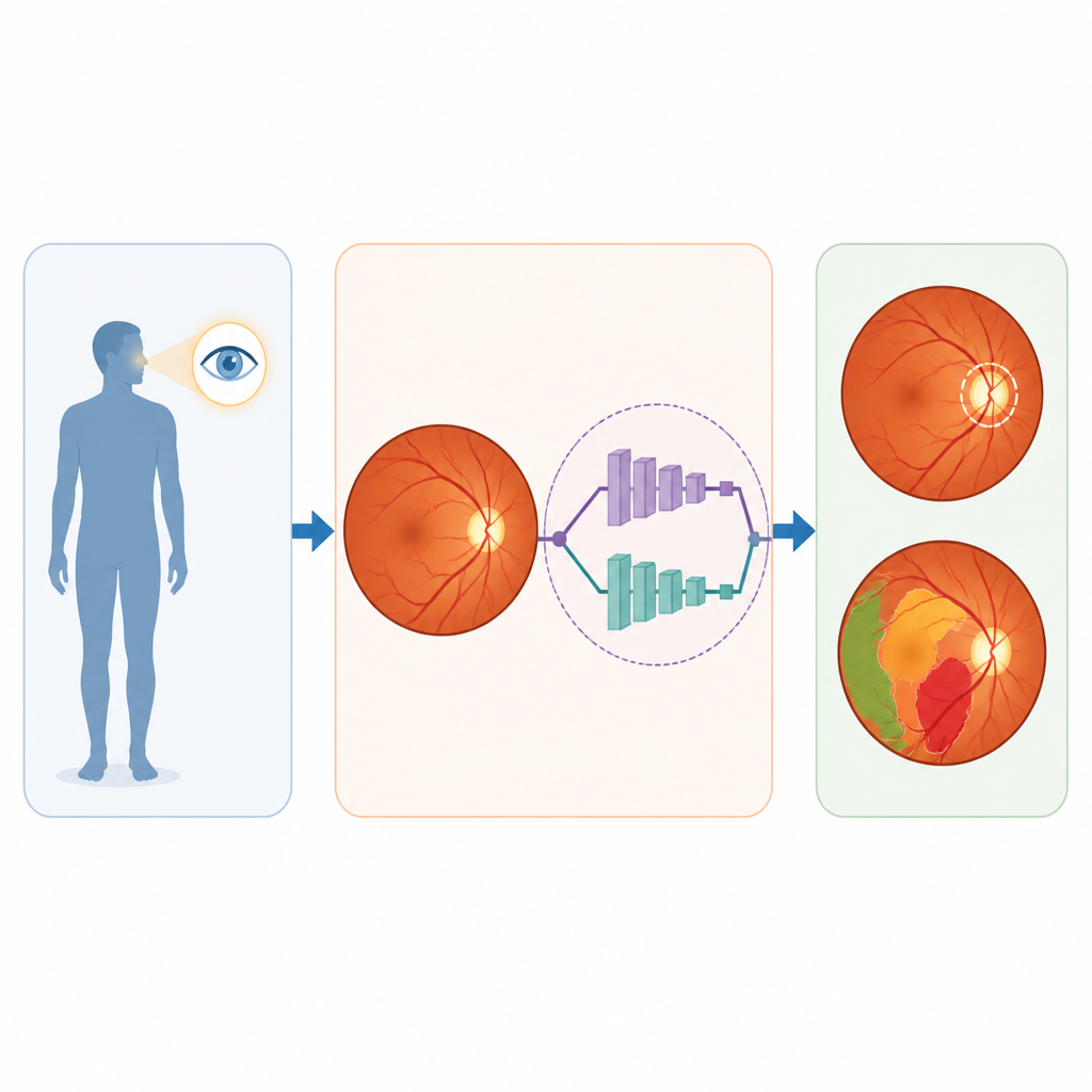
How the smart eye model shares what it learns
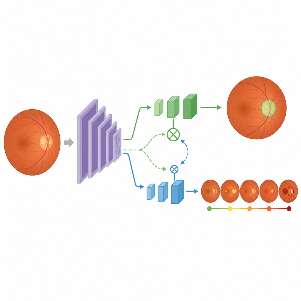
At the heart of GTAM Net is the idea of letting tasks help each other without getting in each other’s way. The system first turns the retinal image into a stack of feature maps that capture shapes, colors, and textures at several scales, from fine vessel details to broader patterns. A special gating unit then decides, for each layer, which parts of this information should be shared and which should be kept private for either drawing the optic disc or grading disease. In parallel, another attention unit lets the two task branches borrow useful hints from one another, so that disease clues can sharpen the disc outline, and knowledge of the disc and other structures can clarify disease grading.
Balancing tasks and working across many datasets
Training such a joint system is tricky, because one task can easily dominate the learning process. To avoid this, the authors let the model estimate how uncertain it is about each task during training and automatically give more or less weight to each objective. They also use a feature pyramid that keeps track of both small details and global layout. GTAM Net is tested on five large public retinal collections that differ in image quality, camera type, and patient mix. On datasets with expert outlines for the optic disc, the system reaches a dice score close to 98 percent, which is on par with or better than earlier disc segmentation tools. For grading diabetic retinopathy, it reports accuracies around 98 to 99 percent on several test sets, outperforming strong existing methods under the same conditions.
Robustness, limits, and what the pictures reveal
The authors go beyond raw scores and examine where the system succeeds or fails. Attention maps show that, when grading disease, the model focuses on suspicious spots such as tiny hemorrhages and bright deposits, while for segmentation it locks onto the disc edge and nearby vessels. When images are blurry, badly lit, or contain rare eye shapes or very heavy bleeding, the outlines and grades can still slip, and errors tend to occur between neighboring severity levels that even specialists find hard to separate. Cross testing, where the model is trained on one dataset and evaluated on another, shows only modest drops in performance, suggesting that the shared, gated design captures general retinal patterns rather than quirks of a single collection.
What this means for future eye screening
In simple terms, the study shows that a carefully designed two in one network can match or surpass separate tools for outlining key eye structures and grading diabetic damage, while staying fast enough for real world screening. By letting the tasks share information in a controlled way and by adjusting their influence during training, GTAM Net delivers accurate, relatively stable performance across varied image sources. While the authors stress that real clinics are more complex than curated test sets and that human judgement remains essential, their results suggest that unified, task aware models could become central building blocks in large scale, automated eye screening programs.
Citation: Sajid, M.Z., Qureshi, I., Hamid, M.F. et al. A gated task-attentive multi-task network for unified retinal image analysis. Sci Rep 16, 16426 (2026). https://doi.org/10.1038/s41598-026-52418-6
Keywords: diabetic retinopathy, retinal imaging, optic disc segmentation, multi task learning, medical AI