Clear Sky Science · en
Deep learning-based automatic field of view planning for prostate MRI in oblique coronal and oblique axial planes
Why this matters for patients and doctors
Prostate MRI has become a key tool for finding and tracking prostate cancer, but image quality still depends heavily on the individual radiographer who sets up each scan. This study asks whether artificial intelligence can take over one crucial setup step so that men, wherever they are scanned, receive more consistent and reliable images without slowing down busy MRI units.

The challenge of aiming the camera
When radiographers plan a prostate MRI, they must decide exactly where to place and angle the scanning window, known as the field of view. Guidelines describe how to tilt this window so that doctors can see the prostate and nearby structures clearly, but in real life this step is time consuming and varies from person to person. Small differences in placement and angle can blur important details, reduce confidence in the results, and sometimes force repeat scans, which cost time and create extra stress for patients.
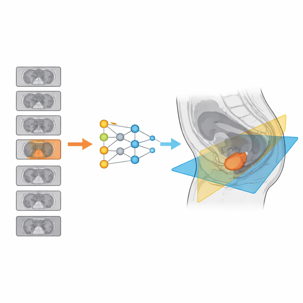
Teaching a computer to plan the view
The researchers built a deep learning system based on a type of image recognition network to handle field of view planning automatically. Using more than 1,400 prostate MRI scans from a public dataset, experts first marked the best slice through the pelvis and drew the ideal scanning windows in two angled directions that are important for prostate imaging. The team then trained their system in two steps: one network learns to pick the most useful slice of the sagittal (side-view) images, and a second network learns to draw the two oblique scan boxes in the right place, size, and angle for that individual patient.
Putting the system to the test
To find out if the computer could match human performance, the authors compared it with experienced readers and with an additional, less experienced rater. They measured differences in slice choice, overlap between the computer and human scan boxes, and how much the angles and positions differed. The system was then tested on 530 new scans from three hospitals that used a mix of scanner models, field strengths, and imaging settings, reflecting the variety seen in real clinics. Across these sites, the overlap between the computer-drawn and expert-drawn fields of view was typically more than four-fifths, and angle differences were only a few degrees, similar to differences seen between human raters.
How useful were the computer plans in practice
Five readers judged whether each computer suggestion was good enough for real diagnostic work, focusing on whether the prostate and seminal vesicles were fully covered. In more than 98 percent of cases overall, the automatically planned scans were rated as clinically acceptable, matching or even exceeding the consistency of less experienced staff. The few failures were mostly linked to very large or unusually shaped prostates, where the gland extended beyond the usual region. The authors suggest that adding more examples of such difficult cases and mimicking common image noise during training could make the system even more robust.
What this could mean for future prostate scans
This multicenter study shows that a deep learning system can take over a key planning step in prostate MRI and reach accuracy similar to skilled radiographers across different hospitals and scanners. By reliably choosing the right slice and drawing well-angled scan windows, the tool could shorten setup time, reduce variation between operators, and help less experienced staff produce high quality images. While the model still needs broader testing on more diverse data and scanners, it points toward a future in which prostate MRI is more standardized, efficient, and consistent for patients everywhere.
Citation: Quinsten, A.S., Wetter, A., Raczkowski, M. et al. Deep learning-based automatic field of view planning for prostate MRI in oblique coronal and oblique axial planes. Sci Rep 16, 14731 (2026). https://doi.org/10.1038/s41598-026-52248-6
Keywords: prostate MRI, deep learning, field of view, medical imaging automation, prostate cancer diagnosis