Clear Sky Science · en
Detection of maxillary sinusitis of endodontic origin in cone-beam CT images using deep learning algorithms
When Tooth Trouble Reaches the Sinuses
Recurring stuffy nose or facial pressure is often blamed on allergies or colds, but a surprising share of sinus problems actually starts with infected back teeth. This study explores how artificial intelligence can read 3D dental scans to spot when sinusitis is linked to dental root infections, helping dentists and doctors reach the right cause more quickly and with greater confidence.
Why Sinus Problems and Teeth Are Connected
The maxillary sinuses sit just above the upper back teeth, separated only by a thin layer of bone. When the soft tissue inside these sinuses becomes inflamed or thickened, people can develop chronic congestion, pain, or a feeling of fullness in the cheek. In many cases a dental infection at the tip of a tooth root irritates the nearby sinus lining, but symptoms may feel like ordinary sinus trouble and the tooth can seem quiet. This makes it hard, even for experienced clinicians, to tell whether sinusitis is driven by dental disease, by other sinus causes, or if the sinus is actually normal.
How 3D Dental Scans Offer a Clearer View
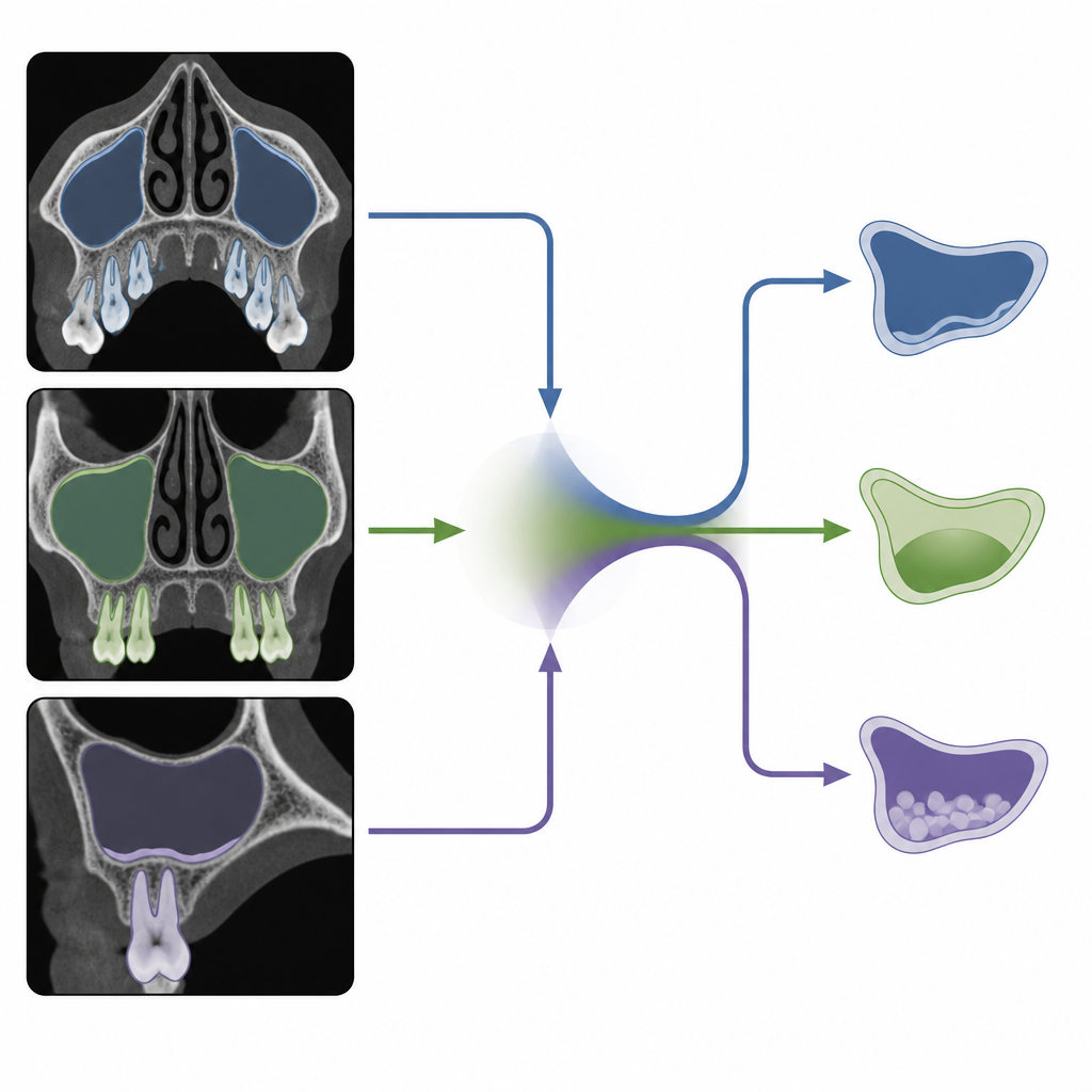
Cone beam computed tomography, or CBCT, is a dental 3D scan that shows both bone and soft tissue in fine detail at a relatively low radiation dose. It can reveal subtle changes around tooth roots and in the sinus lining that are missed on standard medical CT or flat X rays. In this study, researchers gathered 70 CBCT scans from two endodontic clinics and carefully selected over 5,000 two dimensional slices from three viewing angles: top down (axial), front facing (coronal), and side view (sagittal). Expert dentists and an oral radiologist reviewed every scan and agreed on which images showed a normal sinus, which showed sinusitis linked to a diseased tooth root, and which showed sinusitis from other, non dental causes.
Teaching the Computer to Read the Images
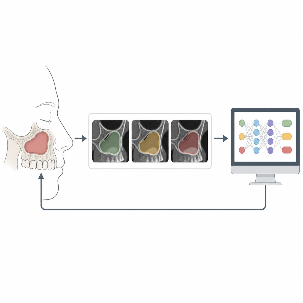
Before training their deep learning system, the team cleaned and enhanced the images. They standardized image size, reduced noise and metal streaks, and sharpened edges so that key structures stood out. They also used a type of generative model to create extra high quality training images, making the data set richer and more varied. The computer pipeline then followed several steps. First, it learned to recognize from each slice which viewing angle it showed. Next, a segmentation stage learned to outline three key regions: tooth roots, the sinus cavity, and the thin mucosal lining. Finally, specialized networks examined each view to extract patterns related to disease, and a “multi view” model combined information from all three angles to decide whether the sinus was normal, affected by a dental root problem, or affected by other sinus disease.
How Well the System Performed
The researchers tested their models on held back images and on a completely separate set of 738 CBCT slices from new patients. The view classifier almost never confused the three scan angles. The segmentation step outlined roots, sinus space, and lining with high overlap compared with expert markings in all three views. Each single view classifier already reached high accuracy, but the multi view network that fused all three perspectives performed best of all. On both the test and external data sets, it correctly labeled every case in the three groups, matching the expert panel’s combined judgment. The authors took several steps to avoid overfitting, including patient wise splitting of data, careful augmentation, and regularization techniques, to make the system more likely to generalize beyond the study sample.
What This Could Mean for Patients
For clinicians, this automated pipeline could serve as a tireless second reader that highlights root related sinus disease, maps the sinus shape, and shows how close infected roots lie to the sinus floor. Such support may help plan root canal therapy, decide how to manage persistent sinus symptoms, and guide implant or sinus lift surgery. While the current study works with 2D slices from a limited field of view and still needs broader testing in more clinics and scanners, it lays a solid foundation for future tools that analyze full 3D scans. In simple terms, the work shows that a carefully built AI can reliably spot when sinus problems are tied to dental roots, offering a clearer path from vague facial pressure to the real source of trouble.
Citation: Sherif, O.A.S., Taha, N.S., Fahmy, A.M. et al. Detection of maxillary sinusitis of endodontic origin in cone-beam CT images using deep learning algorithms. Sci Rep 16, 16254 (2026). https://doi.org/10.1038/s41598-026-52147-w
Keywords: maxillary sinusitis, odontogenic sinusitis, cone beam CT, deep learning, dental imaging