Clear Sky Science · en
Visual information modulates brain network characteristics during static balance following ACL reconstruction – A graph theoretical analysis
Why this matters for everyday movement
Many people who tear a major knee ligament and undergo surgery eventually get back to sports, yet subtle problems can linger for years. This study looks beyond muscles and joints to ask what happens in the brain when people with reconstructed knees try to stand on one leg, with eyes open or closed. Understanding how the brain rewires itself to keep balance could change how we think about rehab, return to sport, and even everyday tasks like walking on uneven ground.
Standing on one leg after knee surgery
The researchers focused on people who had undergone reconstruction of the anterior cruciate ligament (ACL), a key stabilizer inside the knee that is often injured in cutting and pivoting sports. Even long after surgery, many of these individuals report that their knee feels different, and previous work suggests they may rely more on their eyes to stay steady. In this study, 27 people with ACL reconstruction and 24 similar but uninjured volunteers performed a simple task: stand barefoot on one leg for 30 seconds, first with eyes open and then with eyes closed. While they did this, their body sway, knee position, and brain activity were all carefully recorded.
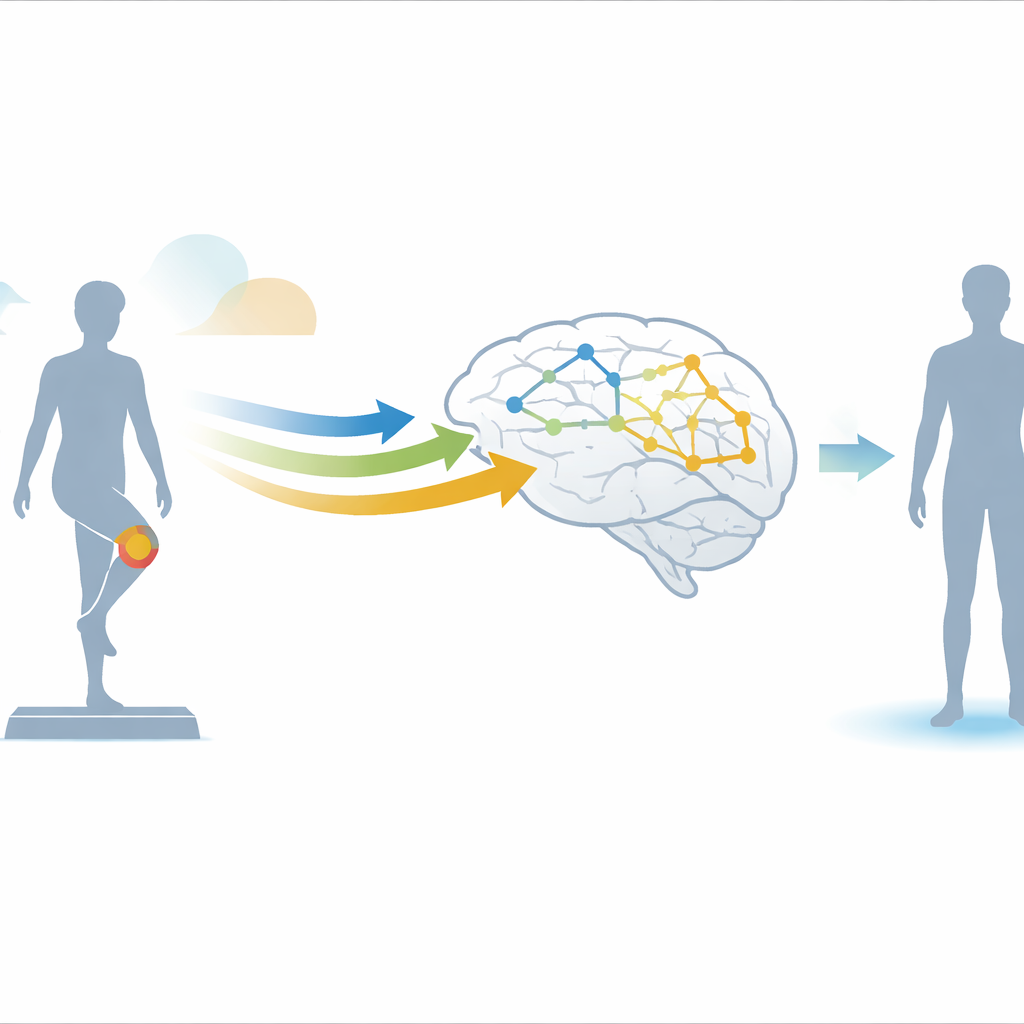
Measuring sway, knee motion, and brain signals
To capture how well participants controlled their balance, the team used a force plate under the standing foot to track tiny shifts in pressure, and a 3D motion-capture system to follow how the body’s center of mass moved over time. From these data they calculated standard measures of sway area and sway speed, as well as the distance between the center of mass and the center of pressure—a combined indicator of how the neuromuscular system keeps the body upright. They also tracked how much the knee was bent on the stance leg, revealing whether people subtly changed their posture to stay steady. At the same time, participants wore a cap with dozens of electrodes that recorded electrical activity from the scalp, allowing the researchers to examine how different parts of the brain coordinated during the balance task.
Seeing balance as a brain-wide network
Rather than just looking for “more” or “less” brain activity, the scientists treated the brain like a network: each electrode was a node, and statistical links between their signals were the connections. Using tools from graph theory, they measured how locally clustered this network was (segregation) and how efficiently information could travel across it (integration). They focused on specific frequency bands of brain rhythms, especially the alpha range, which has been linked to how the brain filters and routes sensory information. Higher clustering in this context suggests that groups of brain areas are working closely together in specialized sub-networks related to the task at hand.
What was different after knee reconstruction
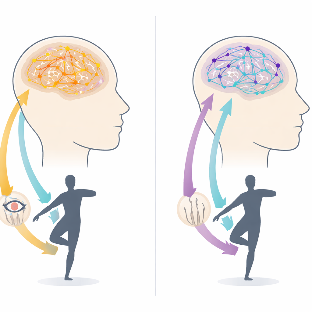
The standout finding appeared only when participants kept their eyes open. In that condition, people with ACL reconstruction showed more tightly clustered brain networks in a low alpha band than uninjured controls, even though their overall sway was similar. This pattern points to more locally specialized processing during balance, hinting that their brains are working harder—yet more efficiently—to organize incoming information. At the same time, the reconstructed leg was held in slightly deeper knee flexion than the other leg in the same person, suggesting a quiet physical adjustment: bending the knee to lower the center of mass and enhance stability. When vision was removed and participants balanced with eyes closed, these brain-network differences vanished, and both groups showed comparable, more challenging sway without clear ACL-related disadvantages.
What this means for real life and rehab
For a layperson, the message is that after ACL reconstruction, the body may look steady, but the brain is doing extra, finely tuned work—especially when vision is available—to keep balance under control. People with a reconstructed knee appear to lean more on visual input and subtle knee bending to achieve the same outward performance as those without injury. When the eyes are closed, everyone must fall back on more automatic body senses, and the advantage of this adapted strategy disappears. These insights suggest that successful rehab is not only about rebuilding muscle and joint stability, but also about how the brain learns to combine sight, body awareness, and movement. Training that challenges balance both with and without vision could help clinicians support more resilient, less visually dependent postural control in athletes returning to sport.
Citation: Grinberg, A., Lehmann, T., Strandberg, J. et al. Visual information modulates brain network characteristics during static balance following ACL reconstruction – A graph theoretical analysis. Sci Rep 16, 14430 (2026). https://doi.org/10.1038/s41598-026-52086-6
Keywords: ACL reconstruction, balance control, brain networks, electroencephalography, sports injury rehabilitation