Clear Sky Science · en
Liver microstructure and biochemical biomarkers in Mormyrus kannume from the River Nile
Why this Nile fish matters
The elephant snout fish of the Nile may look ordinary in a market stall, but inside its body the liver quietly records the story of the river. This organ processes food, stores energy, and helps clear pollution. By carefully mapping what a healthy liver looks like in this species, scientists create a reference guide that future researchers can use to detect early signs of water contamination or disease in one of Africa’s important food fishes. 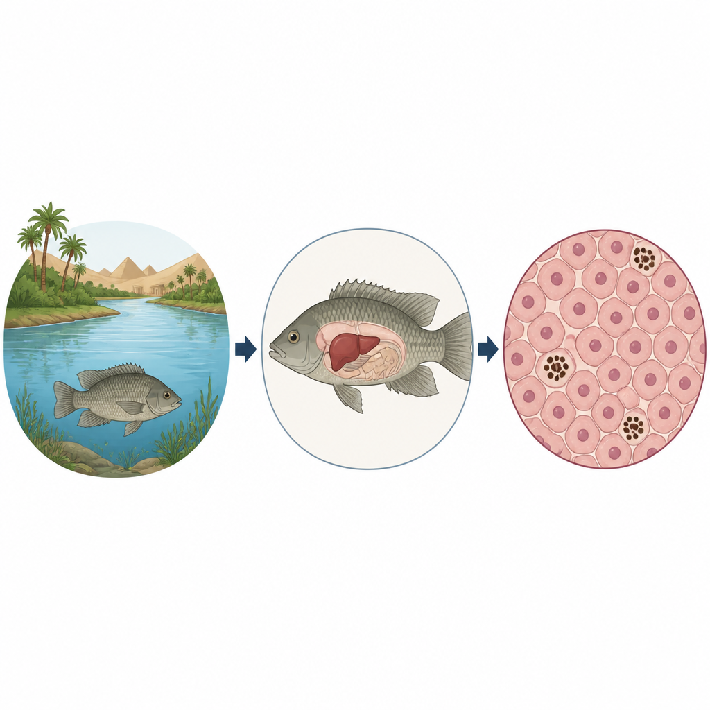
Getting to know a hidden resident
Mormyrus kannume is a nocturnal, carnivorous fish that patrols the bottom of the Nile at night, feeding on insects and small invertebrates. It supports local fisheries and household income along the river. Yet, until now, no one had described the basic liver structure or normal blood chemistry of this species. Without that baseline, it is hard to tell when pollution or other stresses are beginning to harm the fish. The new study set out to fill this gap by examining wild fish from the Nile near Assiut, keeping them briefly under controlled conditions, and then analyzing both their blood and liver tissue.
What the blood can tell us
The team first measured common biochemical markers in the fish’s blood, including glucose, total protein, cholesterol, and several liver enzymes that are often checked in human medical tests. They found that these values fell within ranges reported for other healthy Nile fish, though with some differences tied to diet, activity level, and lifestyle. For example, the elephant snout fish had relatively low glucose levels, which matches its less active, bottom dwelling habits compared with fast swimmers. Cholesterol and protein values also fit with a carnivorous diet. Together, these measurements provide a “normal range” that can later reveal when fish are stressed by low oxygen, handling, or contaminants.
Inside the fish’s internal factory
Looking under the microscope, the researchers saw that the liver cells are arranged in cord like strands radiating around central blood spaces. Between these strands run tiny channels where blood flows, lined with thin endothelial cells and patrolled by Kupffer cells, specialized scavengers that remove debris and invaders. The liver cells themselves are many sided with round nuclei and cytoplasm rich in stored materials. Chemical stains showed that these cells hold abundant glycogen, a storage form of sugar, mainly at their edges. When the scientists digested glycogen with saliva enzymes, the remaining faint staining revealed the structural sugars that support blood vessels. Collagen and elastic fibers formed a delicate framework around major veins and the outer capsule, suggesting a flexible but well supported organ. 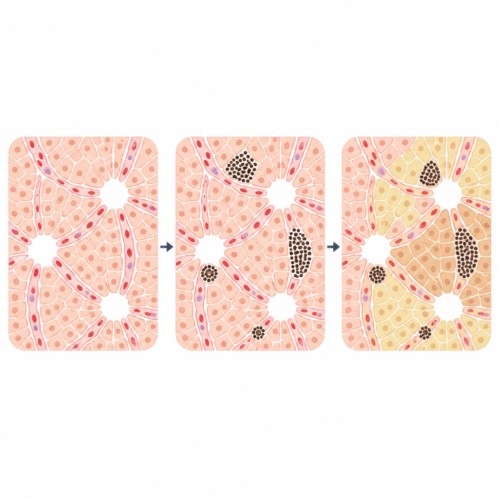
Tiny pigmented sentinels
One of the most striking findings was the variety of melanomacrophage centers, small clusters of pigmented immune cells scattered through the liver. These centers appeared in many shapes, from round to Y shaped and curved forms, and in different locations near veins, blood spaces, and between liver cells. Special stains revealed three pigments: blue toned hemosiderin linked to iron storage and worn out red blood cells; brown lipofuscin related to aging and tissue wear; and dense black melanin, which can help neutralize reactive molecules and support antimicrobial defenses. The pattern of these pigments suggests that the centers are active hubs for recycling iron, clearing damaged material, and responding to environmental stress, making them promising indicators of water quality.
What this means for the Nile and beyond
By combining blood tests with a detailed picture of liver structure and pigment rich immune centers, the study establishes a healthy baseline for Mormyrus kannume. For non specialists, this means that scientists now have a reference atlas for what a normal liver in this Nile fish should look like and how its key blood markers should read. Future surveys can compare new samples to this atlas to detect early liver damage or immune activation linked to pollution, disease, or changing river conditions. In short, the work turns a little known fish organ into a sensitive gauge for the health of the Nile ecosystem and the fisheries that depend on it.
Citation: Ali, A., Abdel-Tawab, H.S., Wassif, E.T. et al. Liver microstructure and biochemical biomarkers in Mormyrus kannume from the River Nile. Sci Rep 16, 15043 (2026). https://doi.org/10.1038/s41598-026-51996-9
Keywords: fish liver, Nile River, biomarkers, melanomacrophage centers, aquatic pollution