Clear Sky Science · en
Integration of resting-state and stimulus-fMRI uncovers reduced network flexibility in post-surgical pain
Why this matters for understanding pain
Surgery can leave people with lingering pain that is hard to explain or treat. This study uses brain scans in rats to explore a simple but important question: does pain after surgery change how flexibly the brain’s networks respond to touch, or does it completely rewire the brain? The answer may help scientists understand why pain sometimes persists and how future treatments might restore healthy brain dynamics.
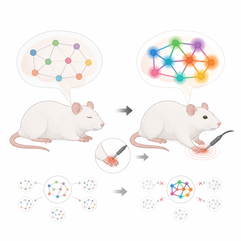
Resting brains and stimulated brains
The researchers combined two types of functional MRI, a brain imaging method that tracks changes in blood flow. One type measured the brain at rest, when no clear task is given. The other measured activity during gentle and stronger pokes to a rat’s hind paw. Some rats had undergone a small surgical cut on the paw to model post-surgical pain, while others had a sham procedure without the cut. By scanning all animals under the same conditions, the team could compare how healthy and pain-affected brains behave both at rest and during touch.
Big-picture wiring stays largely the same
The first surprise was what did not change. Using standard tools that look at how strongly different brain regions are connected overall, the scientists found no clear differences between resting and stimulated scans in either healthy or post-surgery rats. Measures of overall wiring strength and efficiency, such as how easily signals can spread through the network, were stable. Even when they used methods designed to spot changes in specific connections across the whole brain, they still did not see big shifts between rest and stimulation. This suggests that the broad layout of the brain’s communication network remains intact, even in the presence of post-surgical pain.
Zooming in on subtle shifts
To look beyond these global measures, the team turned to more refined analyses that consider many local features of each brain region at once, like how many links it has, how central it is, and how it helps bridge other regions. They used a machine learning method to see whether these combined features could tell healthy and post-surgery networks apart, and whether rest could be distinguished from stimulation. In healthy rats, the analysis clearly separated resting from stimulated states, and different stimulus strengths produced distinct network patterns. In post-surgery rats, these distinctions were still present but much weaker: the networks during rest and stimulation looked more alike in this deeper feature space.
Flexible hotspots versus flat responses
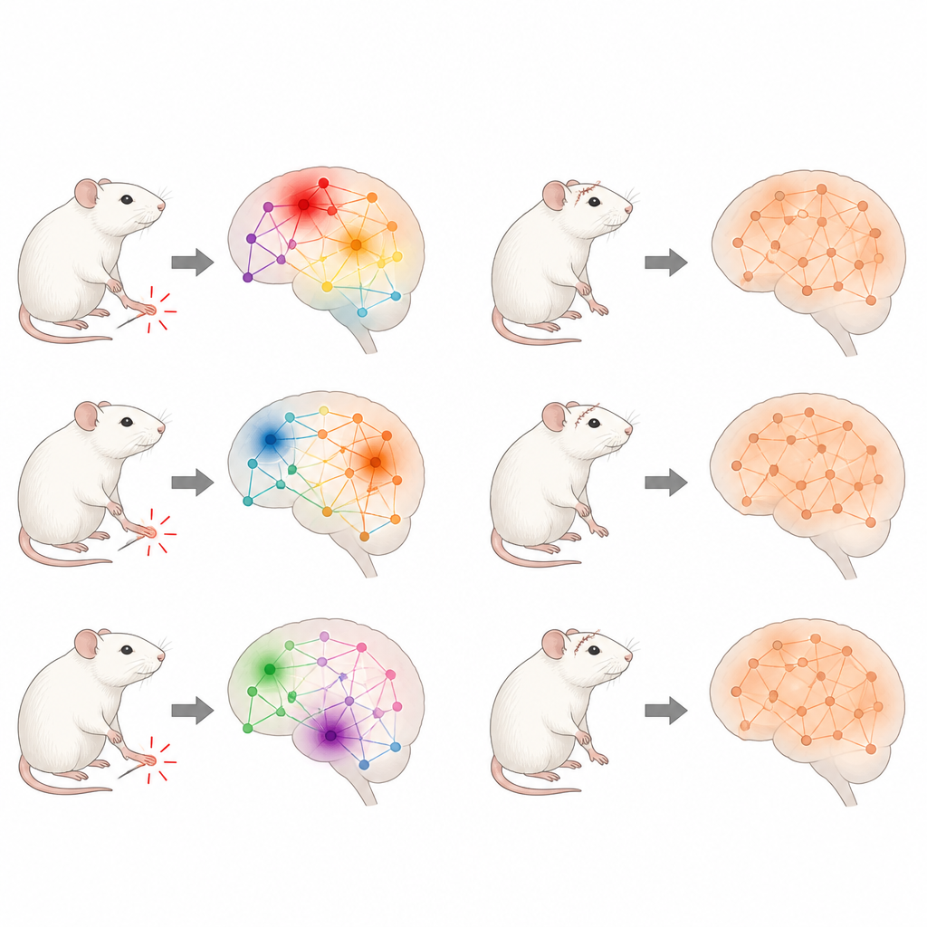
The researchers then asked which specific areas drove these differences. They measured how far each region’s network fingerprint moved when switching from rest to stimulation. In healthy rats, some regions showed especially large shifts, standing out as strong “hotspots” of change, and these hotspots tended to recur across different animals. This produced a spiky pattern in which a few key areas carried most of the adjustment. In post-surgery rats, the pattern was flatter and more spread out. Fewer regions consistently stood out, and the overall changes were more evenly distributed and less varied, pointing to a more rigid and uniform response to touch.
What this tells us about pain and the brain
Altogether, the findings suggest that post-surgical pain does not dramatically remodel the brain’s basic wiring, but it does reduce how flexibly local regions can reconfigure when the body is stimulated. Healthy brains can reshuffle which areas lead the response depending on the situation, while brains in pain show a narrower range of possible patterns. By combining resting and stimulation-based brain scans, this work reveals subtle, pain-related limits in the brain’s ability to adapt that would be missed by looking at resting scans alone.
Citation: Pradier, B., Pogatzki-Zahn, E., Faber, C. et al. Integration of resting-state and stimulus-fMRI uncovers reduced network flexibility in post-surgical pain. Sci Rep 16, 15570 (2026). https://doi.org/10.1038/s41598-026-51946-5
Keywords: post-surgical pain, brain networks, resting-state fMRI, stimulus fMRI, network flexibility