Clear Sky Science · en
Integrating spectral signatures and microbial profiling to differentiate diseased and healthy corals in the Red sea
Why coral color can reveal hidden illness
Coral reefs are the bustling cities of the sea, sheltering a quarter of all marine life and supporting fisheries, tourism, and coastal protection. Yet these underwater metropolises are increasingly struck by mysterious diseases that strip corals of tissue and life. This study explores a new way to check coral health without breaking off pieces for the lab, by reading tiny changes in the colors corals reflect and linking them to the bacteria living on them. The goal is to spot sickness early and gently, before whole reefs begin to crumble.

How coral sickness shows up in color
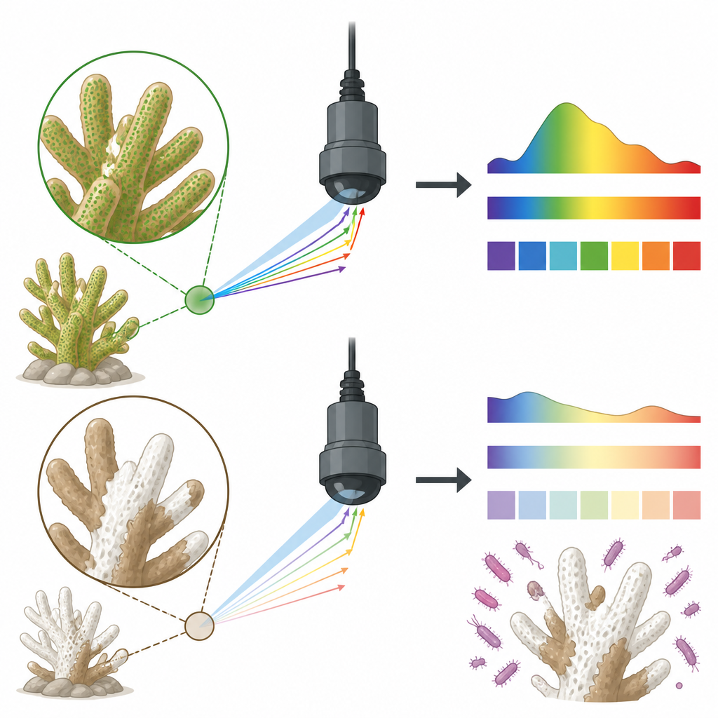
Healthy corals are packed with microscopic algae that give them rich browns and greens and help them turn sunlight into energy. When disease or stress strikes, tissue thins, the white skeleton shows through, and films of microbes can build up on the surface. All of these changes subtly alter how corals reflect sunlight across the rainbow of visible and near infrared light. The researchers used a sensitive underwater instrument to capture these color “fingerprints” from two common Red Sea corals, Acropora humilis and Favia lacuna, carefully comparing healthy colonies with neighbors showing classic white band and white plague symptoms.
Listening to the hidden world of coral bacteria
Corals are more than animals; they are mini ecosystems filled with bacteria that can either support or undermine their health. The team scraped small tissue samples from the same colonies they measured in the water and grew the bacteria in the lab. Using a mass spectrometry system to identify them, they found that healthy corals were dominated by several Bacillus and Cytobacillus species often linked with stable, supportive microbiomes. In contrast, diseased Acropora and Favia were consistently associated with Vibrio species, a group that includes many known marine pathogens. This clear shift from helpful to harmful bacteria provided a biological backdrop for the color changes seen in the field.
Turning coral colors into health clues
With hundreds of closely spaced wavelengths measured from each coral, the challenge was to tease out which parts of the spectrum really mattered. The scientists sharpened the raw reflectance curves using a mathematical trick called a second derivative, which highlights subtle bends in the curve that can be tied to pigments and tissue structure. Diseased corals showed stronger reflectance overall, especially in the green, orange red, and near infrared regions, and displayed distinctive dips at narrow bands between about 450 and 800 nanometers. Healthy colonies, by contrast, kept a stable feature around 675 nanometers, a hallmark of intact chlorophyll from their symbiotic algae. These patterns suggest that disease alters both light absorbing pigments and how light scatters off thinning tissue and exposed skeleton.
Matching light patterns to microscopic partners
To see whether bacterial communities and light patterns truly traveled together, the team combined the spectral data with the bacterial profiles using clustering and other statistical tools. Corals and bacteria grouped into distinct sets whose color signatures lined up with health status rather than water conditions like temperature, saltiness, or pH, which were similar across sites. Certain wavelength ranges around 450, 500, 600, 700, and 800 nanometers stood out as especially good at separating healthy from diseased colonies. Even when the same coral species was studied at different reefs, these narrow bands repeatedly captured the shift from symbiont rich, stable tissue to damaged, microbe laden surfaces.
What this means for protecting reefs
The study shows that it is possible to distinguish healthy and diseased Red Sea corals by reading their spectral signatures and tying those signals to changes in their bacterial partners. Although based on a modest number of samples and a few coral species, the work points to a future in which divers, drones, or satellites may scan reefs for specific “problem colors” that hint at early disease, without taking a single fragment. By identifying a short list of key wavelengths and linking them to disease associated bacteria, the authors lay the groundwork for non invasive tools that could help reef managers detect trouble sooner and better understand how microbes tip the balance between coral health and decline.
Citation: Khalifa, A.M., ElBaghdady, K.Z., Hamed, M.M. et al. Integrating spectral signatures and microbial profiling to differentiate diseased and healthy corals in the Red sea. Sci Rep 16, 15349 (2026). https://doi.org/10.1038/s41598-026-50675-z
Keywords: coral disease, hyperspectral sensing, coral microbiome, Red Sea reefs, marine monitoring