Clear Sky Science · en
Veterinary fracture diagnosis: a deep learning model for dogs long bone fractures
Why this matters for pets and their people
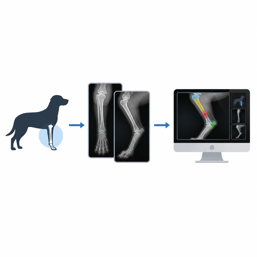
When a dog suddenly starts limping, every minute waiting for answers feels long. Vets rely on X ray images to decide whether a bone is broken and how serious the damage is, but reading these images can be slow and sometimes uncertain. This study explores how computer programs that learn from examples can help veterinarians spot and classify certain leg fractures in dogs more quickly and consistently, offering faster guidance for treatment and peace of mind for owners.
Broken bones in everyday vet practice
Fractures in dogs are a routine problem in veterinary clinics. Affected animals may show pain, swelling, trouble standing, or an oddly angled limb. Long bones in the legs, such as the femur or tibia, are especially important because they carry the dog’s weight. These breaks come in many patterns, from clean straight lines to twisted or crushed pieces, and vets must judge where the break is, how the pieces lie, and whether a joint is involved. Getting this right is crucial because it shapes the choice between simple rest, splints, or complex surgery.
How smart software reads X rays
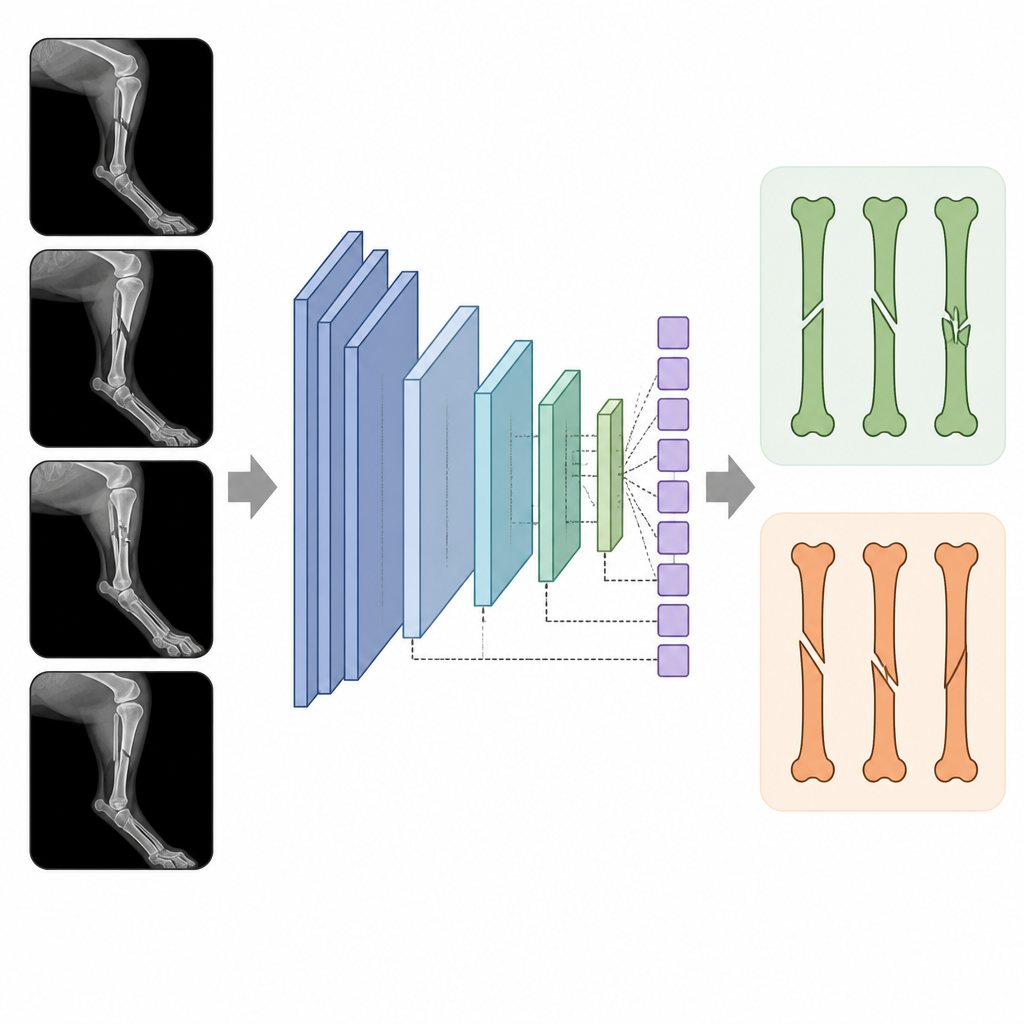
The authors built a computer system that looks at standard X ray images of dog legs and decides between two common long bone fracture types called oblique and overriding. The core of their system is a powerful image recognition model known as ResNet50, a form of deep learning that can pick up subtle patterns in pictures. To compare its performance, they also tested several other widely used models. Because there are few labeled veterinary images available, they added an automatic step that isolates only the part of the X ray that contains the fracture, helping the model focus on what matters most.
Making the most of a small image collection
One challenge in this work is that the researchers began with only 44 suitable X ray images, far fewer than deep learning systems usually require. To get around this, they used a strategy called data augmentation, in which the computer creates many realistic variations of each original image by rotating, zooming, and slightly altering it while keeping the fracture pattern intact. They also carefully balanced the number of images in each fracture group so the model would not favor one type over the other. The dataset was then split into training, validation, and test sets so that the final results would reflect performance on images the system had not seen before.
How well the system performed
After training, the ResNet50 model almost never misclassified a fracture in the test images. It reached an accuracy close to 100 percent and scored very highly on related measures that capture how often it correctly identifies and correctly rejects each fracture type. When compared with six other deep learning models, ResNet50 not only classified fractures more accurately but also showed a strong ability to separate the two categories across many test cases. The authors further examined how quickly each model runs and how much computer memory it uses, finding that ResNet50 struck a favorable balance between speed, resource use, and reliability.
What this means for future vet visits
This study suggests that carefully designed learning systems can support veterinarians by highlighting and classifying certain dog leg fractures on X rays with very high consistency. While the current work focuses on two fracture types and a relatively small image set, the same approach could be expanded to more complex injuries and larger collections of images. In time, such tools may act as a quiet assistant in the clinic, offering a rapid second opinion that helps vets plan treatment sooner and gives injured dogs a better chance at quick and accurate care.
Citation: Saber, A.S., Selim, I., Askr, H. et al. Veterinary fracture diagnosis: a deep learning model for dogs long bone fractures. Sci Rep 16, 15098 (2026). https://doi.org/10.1038/s41598-026-50387-4
Keywords: dog bone fractures, veterinary radiology, deep learning, X ray imaging, fracture diagnosis