Clear Sky Science · en
Comparative study of pathology of various organs of rhesus macaques exposed to two different doses of acute total-body radiation
Why radiation injuries matter to everyday life
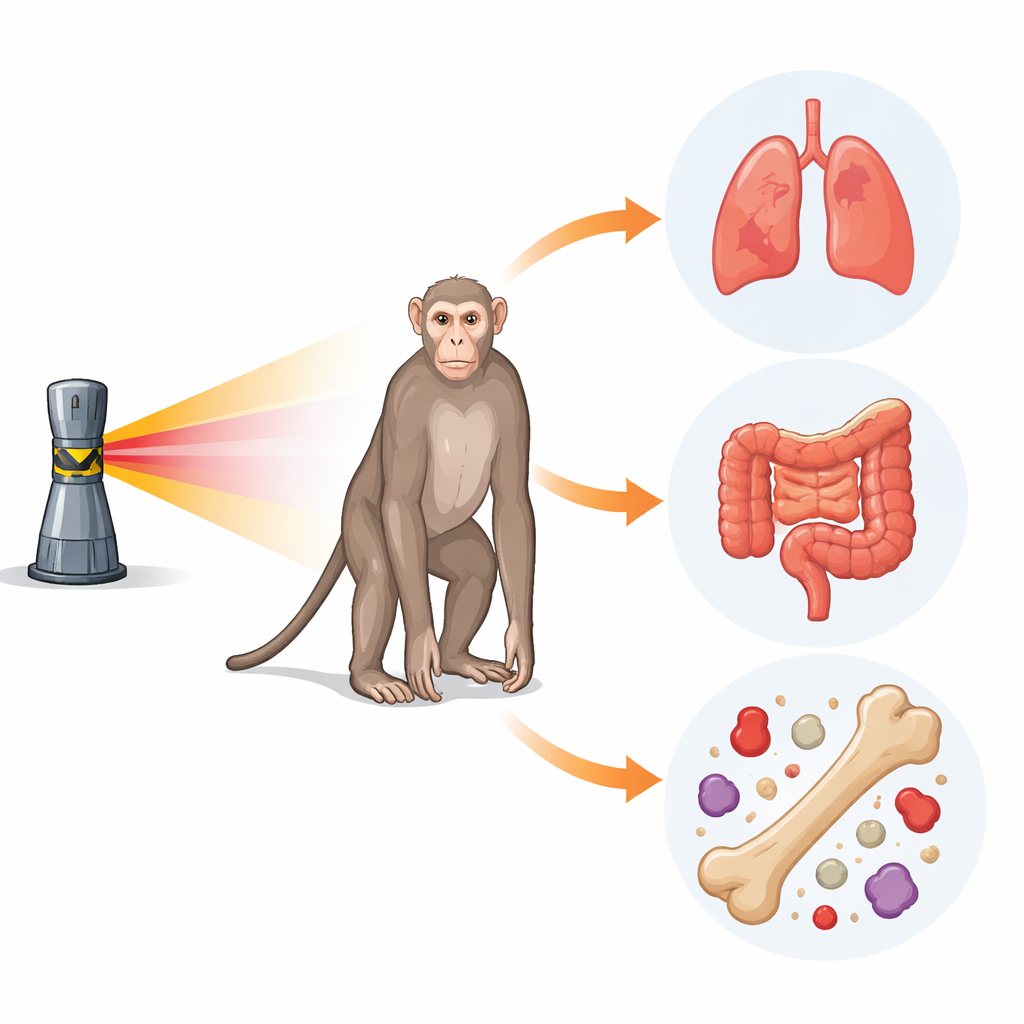
In a world where nuclear power plants, medical radiation devices, and even dirty bombs are real possibilities, understanding what high doses of radiation do inside the body is more than an academic question. This study used rhesus macaques—monkeys that closely resemble humans—to trace how a single, intense blast of whole-body radiation ripples through major organs over two months. The work helps doctors and regulators design and test drugs that could one day save lives after a nuclear accident or attack.
Looking inside the body after a powerful blast
The researchers exposed 31 male and female rhesus macaques to one of two near‑lethal levels of cobalt‑60 gamma radiation, roughly comparable to doses that would cause acute radiation sickness in people. After exposure, the animals received careful supportive care much like human patients would—fluids, antibiotics, and symptom relief—while their health was tracked for 60 days. At the end of the study, or when animals became too sick to recover, the team performed detailed tissue examinations under the microscope and measured changes in blood counts and blood chemistry. This allowed them to connect what was happening in the bloodstream with the damage seen in specific organs.
Blood and immune cells take the hardest hit
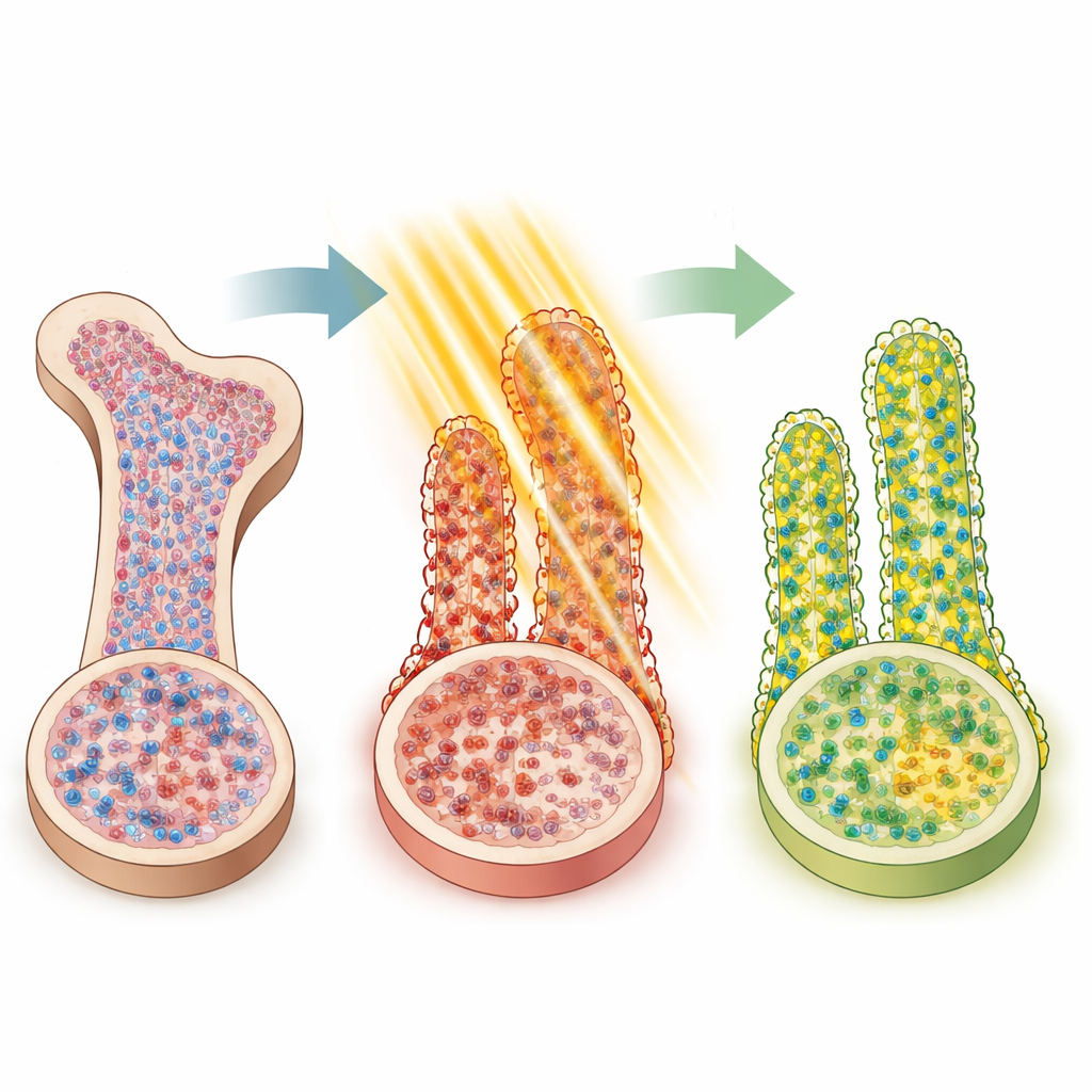
Because radiation especially harms rapidly dividing cells, the blood‑forming system in the bone marrow was a major target. White blood cells, which fight infection, plummeted more than 28‑fold within two weeks. Platelets, needed for clotting, also dropped sharply, and some animals at the higher dose developed severe platelet shortages. Red blood cells fell more slowly but remained depressed for weeks. Under the microscope, bone marrow in the breastbone and immune tissue in the spleen showed dramatic loss of cells, especially in animals that did not survive. Interestingly, although the higher dose generally caused worse blood shortages, one puzzling pattern emerged: the lower dose sometimes showed more visible depletion in bone marrow sections, likely because animals at the higher dose died before the full tissue changes could develop.
Gut and lungs reveal hidden internal damage
The lining of the small intestine, another tissue that renews itself rapidly, also showed clear injury. In many animals, finger‑like villi that absorb nutrients were shortened, fused, or lost, and the deep pockets where new cells are born were disrupted. Damage was usually worse at the higher radiation dose and often more pronounced in females. In the large intestine, the tiny glands that produce mucus and help maintain the barrier to germs were partly destroyed, especially at 6.5 gray. The lungs, however, were the most consistently dose‑dependent organ: higher‑dose animals showed thicker, damaged air‑sac walls and fluid buildup, signs that the delicate surfaces needed for gas exchange were compromised. In contrast, the heart, kidneys, liver, and bladder showed mostly mild or subtle changes, suggesting that at these doses they are less critical to short‑term survival than the blood, gut, and lungs.
Sex, dose, and the body’s attempt to recover
By following animals over time, the team also saw how the body fights back. After deep early crashes, many blood cell types rebounded, sometimes overshooting their original levels by day 60, especially in animals at the higher dose. This overshoot hints at powerful repair programs kicking in once enough stem cells survive to restart production. Blood chemistry tests showed shifting markers of kidney, liver, and general tissue stress, such as changes in creatinine, bilirubin, lipids, and an injury enzyme called LDH, again more disturbed at the higher dose. When the scientists compared males and females, they found that sex did not strongly change whether animals survived, but it did shape how and when specific tissues and blood cell types recovered, particularly in the gut and blood‑forming organs.
What this means for protecting people
For a general reader, the key message is that a single strong dose of radiation does not harm the body in a uniform way. Instead, certain organs—the bone marrow, immune tissues, gut, and lungs—are much more vulnerable, and the pattern of damage and recovery depends on both how much radiation is received and whether the individual is male or female. By mapping these organ‑by‑organ responses in an animal model that closely mirrors humans, this study provides a roadmap for developing and testing future drugs that could protect or repair the most sensitive systems after a nuclear event, and it shows regulators which signs to watch when judging whether a new treatment truly works.
Citation: Brink, M.W., Petrus, S.A., Carpenter, A.D. et al. Comparative study of pathology of various organs of rhesus macaques exposed to two different doses of acute total-body radiation. Sci Rep 16, 14034 (2026). https://doi.org/10.1038/s41598-026-49844-x
Keywords: acute radiation syndrome, total body irradiation, rhesus macaque, multi-organ injury, radiation countermeasures