Clear Sky Science · en
Comparing DNA metabarcoding with light microscopy to identify eukaryotic phytoplankton in the Baltic Sea, Kattegat and Skagerrak
Why tiny sea plants matter
Across the Baltic Sea and its neighboring waters, vast communities of microscopic plants called phytoplankton fuel marine food webs and influence water quality. Keeping track of which species are present and how they change over time is essential for spotting harmful blooms, understanding climate impacts, and managing fisheries. This study asks whether a modern DNA based technique can complement the long standing microscope based surveys that many monitoring programs still rely on.
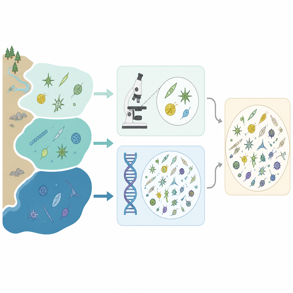
Two different lenses on the same community
For decades, specialists have identified phytoplankton by looking at preserved water samples under light microscopes, carefully counting cells and matching shapes to species. This approach provides direct visual evidence but demands expert time and can miss very small or delicate cells. DNA metabarcoding offers a different route. By filtering seawater, extracting all the genetic material, and sequencing a marker gene shared across many organisms, researchers can infer which taxa are present from their DNA signatures, even if the cells are too small or similar looking to distinguish by eye.
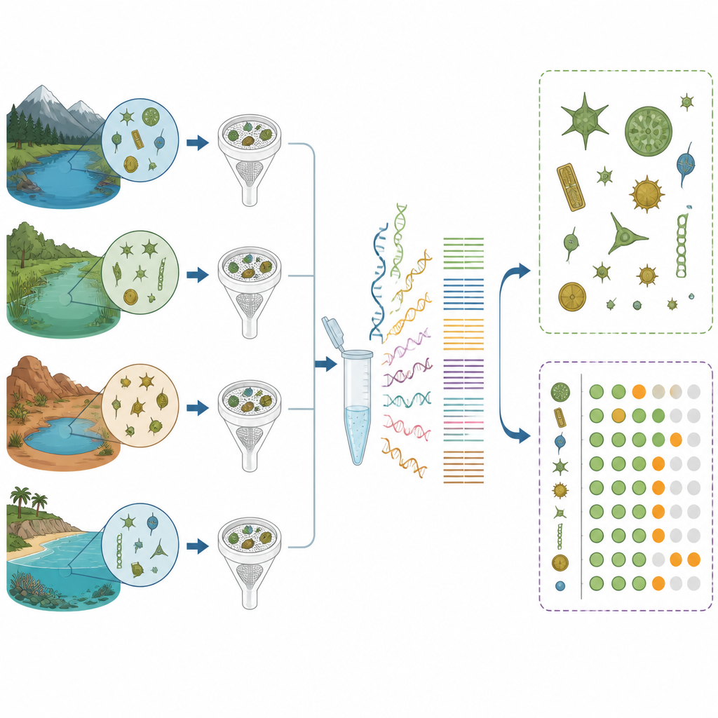
A natural testbed with changing salt levels
The Baltic Sea, Kattegat, and Skagerrak form a connected system with a strong gradient in saltiness, ranging from almost fresh water in the northern Bothnian Bay to fully marine conditions in the Skagerrak. This variety hosts a mix of freshwater and marine phytoplankton and makes identification by shape alone particularly demanding. The team collected 232 surface water samples at 17 stations along this gradient between early 2019 and early 2020. Each sample was examined in two ways: by the traditional Utermöhl microscope method and by DNA metabarcoding of the 18S ribosomal RNA gene, a standard marker for eukaryotic microbes.
What DNA reveals that microscopes miss
Overall, DNA metabarcoding detected many more orders, genera, and species than microscopy. For example, it picked up numerous small celled taxa that are difficult or impossible to recognize morphologically, such as several tiny dinoflagellates and haptophytes. The two methods agreed on the presence of 43 percent of the most common genera, but each also found groups that the other missed. Some species appeared only in microscope counts, often because the necessary DNA reference sequences are still lacking or incomplete in public databases. Others appeared only in the DNA data, especially among very small organisms that tend to be lumped into broad categories under the microscope.
Counting cells versus weighing biomass
The researchers also asked whether DNA sequence counts could stand in for actual abundances. They tried several ways to normalize the sequence data, including using added synthetic DNA as a reference and adjusting by the total DNA concentration. When they compared these measures to microscopic estimates of cell numbers, cell size (biovolume), and carbon content, the matches were generally weak and varied among taxonomic groups and regions. Interestingly, DNA results aligned better with carbon and biovolume than with simple cell counts, suggesting that gene copy number may scale more with cell size than with the number of individuals. Even so, no normalization approach reliably translated DNA reads into absolute biomass across the whole dataset.
How the two methods work together
Despite the quantitative limits, DNA metabarcoding proved more reproducible than microscopy for several major phytoplankton classes and captured clear regional patterns in community composition along the salinity gradient. It also highlighted potentially harmful bloom forming groups that are often hard to pinpoint in routine counts. The authors conclude that DNA based surveys are not yet ready to replace microscopes in long term monitoring, especially where precise biomass estimates and species level identifications are needed. However, as reference databases grow, long read sequencing becomes more common, and scientists better understand how gene copy numbers vary among taxa, metabarcoding can greatly extend biodiversity assessments. Used alongside traditional microscopy, it offers a powerful way to see both the familiar and the hidden parts of marine phytoplankton communities.
Citation: Torstensson, A., Brugel, S., Andersson, A.F. et al. Comparing DNA metabarcoding with light microscopy to identify eukaryotic phytoplankton in the Baltic Sea, Kattegat and Skagerrak. Sci Rep 16, 15743 (2026). https://doi.org/10.1038/s41598-026-48838-z
Keywords: phytoplankton, DNA metabarcoding, Baltic Sea, marine monitoring, microscopy