Clear Sky Science · en
Precise ablation of cholesteatoma using a 445-nm diode laser
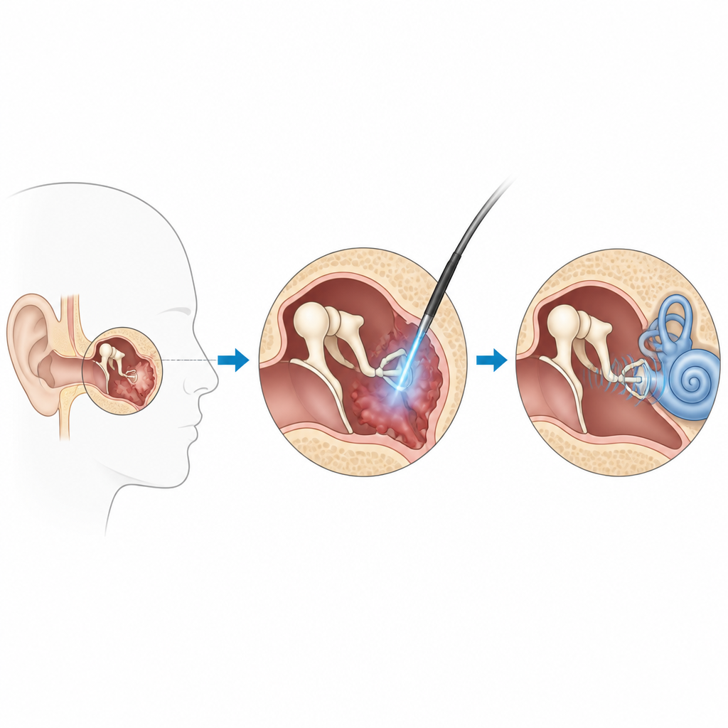
Gentle surgery for a hidden ear problem
Many people live for years with a slow, silent ear disease called cholesteatoma before it is discovered, often after it has already damaged the tiny bones that carry sound. Treating it requires delicate surgery deep inside the middle ear, where even a slip of a fraction of a millimeter can affect hearing. This study explores whether a specific blue laser can help surgeons remove the diseased tissue more precisely while leaving the fragile hearing bones intact.
Why tiny ear bones need careful treatment
Cholesteatoma is a growth of skin-like tissue that creeps into the middle ear and gradually eats away at nearby bone. The three middle ear bones, each only a few millimeters in size or less, form a chain that passes sound from the eardrum to the inner ear. If these bones are harmed during surgery, patients can lose more hearing even after the disease is removed. Surgeons already use different kinds of lasers to help cut and seal tissue, but each type has tradeoffs in terms of penetration depth, bleeding control, and ease of use in the cramped space of the middle ear.
A blue laser tuned for delicate work
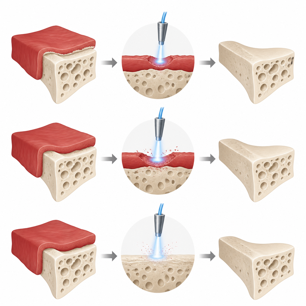
The researchers focused on a diode laser that emits blue light at 445 nanometers. This light is strongly absorbed by blood and soft tissue, but less by hard bone. The team asked whether short, single pulses of this laser could shave away thin layers of diseased tissue without biting into the underlying bone. They first built a model using pig ear cartilage covered by a thin soft layer to imitate human middle ear bones coated with cholesteatoma. They then tested different laser powers, distances between the fiber tip and tissue, and angles of approach, measuring how deep each pulse cut using high-resolution imaging and microscopic analysis.
How the laser behaves in real tissue
In the pig model and in donated human cholesteatoma samples, the laser removed only the top few hundred micrometers of soft tissue when fired in 100 millisecond pulses. As the power increased from low to higher settings, the depth of removal increased in a nearly straight-line fashion, but even at the highest power the cuts stayed shallower than the thinnest vulnerable parts of the ear bones. Importantly, the underlying cartilage remained structurally intact, with clear boundaries between the removed layer and preserved deeper tissue. Changing the distance and angle of the laser fiber affected how deep the cuts were, but within realistic hand-held ranges these variations stayed within a safety margin of a few tenths of a millimeter.
Selective removal of disease while sparing bone
To mimic real surgery, the team also applied overlapping pulses to create continuous paths of removal. These runs produced even, shallow channels that stripped away the soft layer but did not harm the supporting cartilage. When they moved on to human ear bones and cholesteatoma tissue, the difference became even clearer: at low power, the blue laser reliably ablated the soft cholesteatoma but left the bones essentially untouched. Only at the highest tested power did the bones show limited, shallow damage, while the diseased tissue showed much more pronounced removal. This reflects the way blue light is more readily absorbed in blood-rich soft tissue than in dense mineralized bone.
What this means for ear surgery and hearing
The findings suggest that a 445 nanometer blue diode laser, used in short single pulses, can help surgeons peel away cholesteatoma layer by layer while keeping the delicate hearing bones structurally safe. Because the cutting depth is predictable and remains below critical bone thicknesses, the laser offers a fine level of control that is well suited to the tight confines of the middle ear. While further clinical work is needed, this approach could support hearing-preserving surgery by combining effective removal of diseased tissue with protection of the tiny structures that make hearing possible.
Citation: Enzian, P., Detje, A.M., Lange, B. et al. Precise ablation of cholesteatoma using a 445-nm diode laser. Sci Rep 16, 15995 (2026). https://doi.org/10.1038/s41598-026-47908-6
Keywords: cholesteatoma, middle ear surgery, blue diode laser, ossicle preservation, laser ablation