Clear Sky Science · en
Multi-scale covariance filtering for edge-preserving medical image fusion
Sharper brain scans from blended views
Doctors often look at several kinds of brain scans to understand what is happening inside the skull, but each scan type shows only part of the story. This study introduces a new way to combine different medical images into a single, clearer picture that keeps fine details at the edges of important structures, helping clinicians see both soft tissue and bone more precisely.
Why mixing scans can help patients
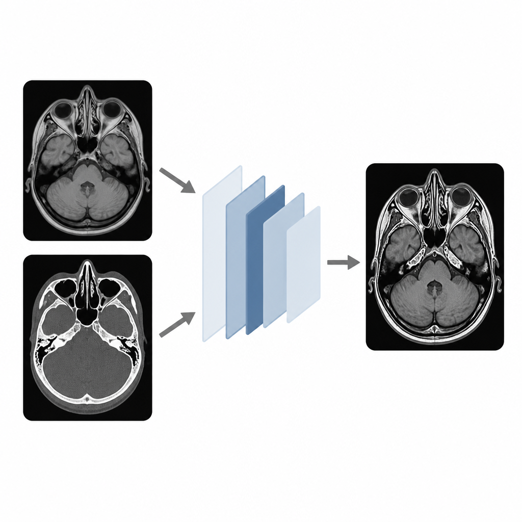
Modern hospitals routinely capture brain images using tools like CT and MRI, each highlighting different aspects of anatomy and disease. CT excels at showing dense structures such as bone, while MRI is better for soft tissues, fluid, and lesions. Looking at these images side by side can be time consuming and can make subtle problems easy to miss. If the information from multiple scans can be fused into one well balanced image, doctors may spot abnormalities more confidently and plan treatments, such as surgery or radiotherapy, with greater accuracy.
The problem with earlier fusion methods
Earlier techniques for fusing medical images often relied on mathematical filters or deep learning systems that either smoothed away delicate structures or introduced visual artifacts along tissue boundaries. Some methods blurred the contrast between gray and white matter, while others created halos around the edges of the skull or ventricles. Many approaches also demanded heavy computation or complex training, which limits their use in busy clinical settings. Radiologists need fused images that preserve natural edges, maintain realistic brightness and contrast, and can be produced quickly from standard scans.
A new recipe for clearer combined images
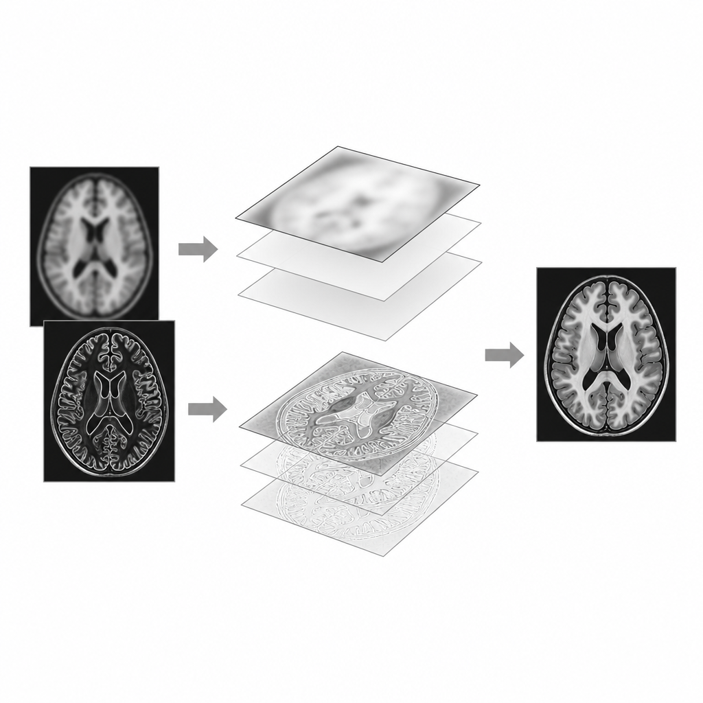
The authors propose a fusion framework built around a two step view of each image: a base layer containing broad brightness patterns and a detail layer containing edges and textures. First, every input scan is gently smoothed to capture overall shading, then the fine details are isolated by subtracting this smoothed version from the original. Next, a saliency process looks for pixels that stand out locally, such as sharp borders between tissues, by applying special edge highlighting and soft blurring filters. This produces early “weight maps” that indicate which scan carries the most useful information at each location, but these maps can be noisy or blocky if used as they are.
Letting local statistics refine the blend
To clean up these weight maps and keep edges crisp, the method uses what the authors call covariance filtering. Instead of borrowing guidance from an extra image, this step examines how the brightness of each scan and its weight map vary together in small neighborhoods. Where there is a strong local relationship, the weights are adjusted to follow the structure of the underlying anatomy, smoothing out flat regions while preserving sharp boundaries. Separate refined weights are produced for the base and detail layers and then normalized so that all scans contribute in a balanced way. Finally, the base and detail layers from all images are recombined using these refined weights to form a single fused image that reflects both global shading and fine structure.
How well the method performs
The researchers tested their approach on three sets of paired brain scans that included different combinations of CT, T1 weighted MRI, T2 weighted MRI, and contrast enhanced MRI. They compared the results with a wide range of existing fusion methods using standard measures of brightness, contrast, sharpness, information content, and edge preservation. Visually, their fused images showed clear ventricles, well defined skull outlines, and improved separation of gray and white matter without halos or over smoothing. Quantitatively, the method matched or exceeded competing techniques on key indicators, especially those linked to edge quality and detail retention, while keeping the computational cost relatively low.
What this means for future medical imaging
In simple terms, the study shows that carefully guided blending of scans, using local statistics rather than heavy learning models, can produce single brain images that look more natural and contain more clinically useful detail. By preserving sharp anatomical boundaries and balancing the strengths of CT and MRI, this fusion method could support more confident diagnosis and planning in neurology and oncology. The authors note that the same idea can be extended to three dimensional image stacks and integrated with smarter region selection, potentially making multi scan fusion a routine tool in precision medicine.
Citation: Sharma, S., Rani, S., Dogra, A. et al. Multi-scale covariance filtering for edge-preserving medical image fusion. Sci Rep 16, 16177 (2026). https://doi.org/10.1038/s41598-026-47798-8
Keywords: medical image fusion, brain imaging, MRI CT fusion, edge preservation, diagnostic imaging