Clear Sky Science · en
Intravascular ultrasound wall shear stress imaging in stented coronary arteries with ultrafast Doppler
Why this matters for heart stents
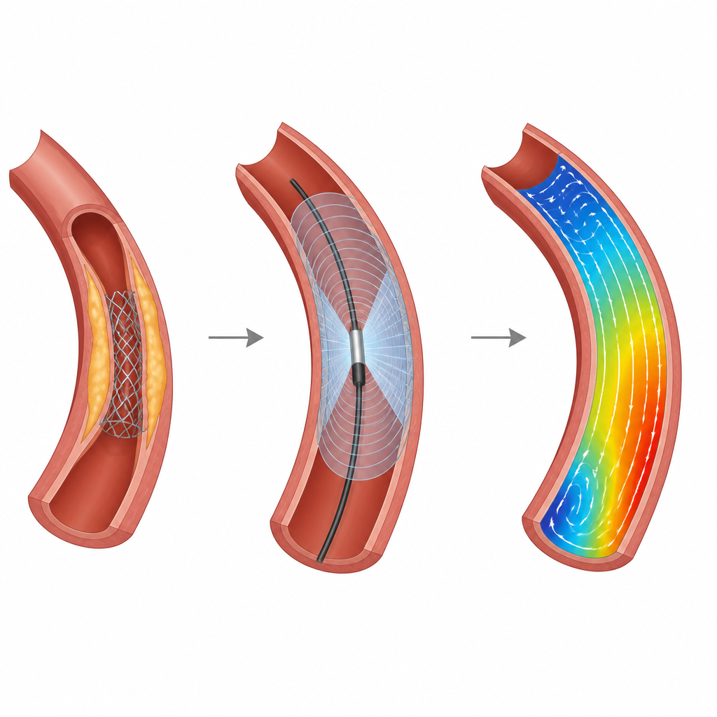
Millions of people each year receive tiny metal tubes called stents to prop open clogged heart arteries. While these devices save lives, some stents slowly clog again over time. This study explores whether doctors could one day watch how blood actually flows around a new stent, in real time, using ultrasound from inside the artery. That information could help them fine tune stent placement and better spot patients at risk for future trouble.
Blood flow and the hidden force on artery walls
When blood flows through an artery, it drags along the vessel wall, creating a gentle frictional force. Scientists call this wall shear stress, but it can be thought of as the rubbing of blood against the artery lining. If this rubbing is too low or too uneven, the artery wall is more likely to develop fatty buildup or react badly to a stent. Studies have shown that areas with disturbed rubbing are linked to plaque growth and to renewed narrowings after a stent is placed.
Limits of current views inside the heart
Today, cardiologists mainly rely on X ray movies and special cameras threaded into the artery to see how a stent is sitting. These tools are excellent at showing the shape of the vessel and whether the stent looks fully opened and pressed against the wall. However, they cannot directly show how blood is moving around the metal struts, or whether the rubbing on the wall has become dangerously low or high. To estimate that, researchers usually build computer models from scans, a process that is accurate but slow and impractical for guiding a live procedure.
A new way to watch blood flow from inside the vessel
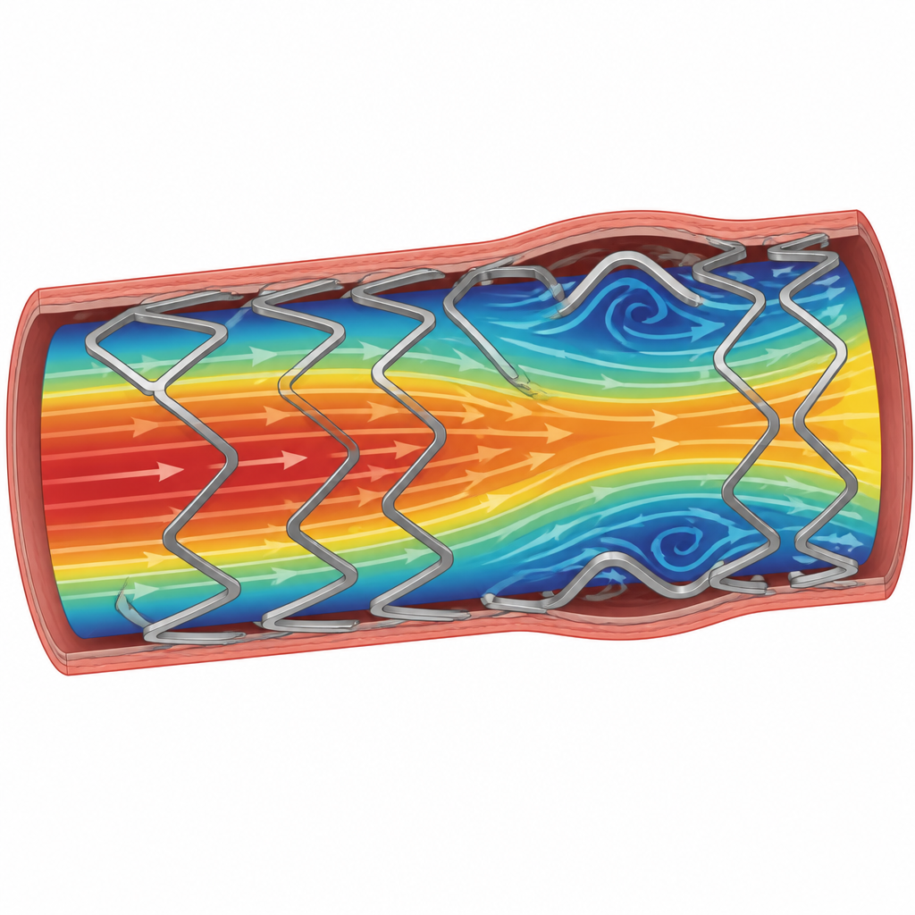
The team behind this study tested an intravascular ultrasound method that does more than draw a picture of the artery. Using very fast pulses and advanced processing, their system measures the speed and direction of blood at many points inside the vessel. From these detailed flow maps, they can calculate how strongly blood is rubbing the wall along the length of the artery, even when a stent is in place. They built realistic model arteries, including one based on an actual patient scan, and created versions with no stent, a partly expanded stent, and a fully expanded stent. They then compared the ultrasound results with high end computer simulations built from micro CT images of the same models.
How well the technique performed
Across all of the model arteries, the ultrasound based flow maps were generally close to the computer simulations. On average, the difference in measured blood speed was about 13 percent, and the patterns in space matched well. In straight, fully expanded stents, the agreement was especially strong. In more twisted or narrowed vessels, and in cases where the stent was not fully pressed against the wall, differences grew but the main trends still lined up. Importantly, when the stent was left partly expanded and lifted from the wall, the ultrasound maps showed lower average rubbing in those regions and more patchy variation, features that have been linked to a higher chance of the stent clogging again.
First steps toward full 3D flow imaging
Beyond flat cross section views, the researchers also tried a tiny forward looking ultrasound array designed to sit inside a coronary artery. With this device, they produced the first real time three dimensional flow and rubbing maps in simple model vessels. These early tests showed the expected pattern of fastest flow and highest rubbing at and just beyond a narrowing, hinting that truly volumetric imaging during a real procedure may be within reach.
What this could mean for patients
This work suggests that a future catheter might let cardiologists not only see where a stent sits but also how blood glides around it. By identifying regions where the blood rubbing on the artery wall is too low or too uneven, they could adjust stent expansion on the spot, schedule earlier checkups for higher risk patients, or feed the data into long term digital models of a person’s coronary health. While the current study used laboratory models and steady flow, it provides an important proof of concept that intravascular ultrasound alone can capture the key forces that influence whether a stent will stay open or narrow again.
Citation: Singh, T.C., Strassle Rojas, S., Shah, I. et al. Intravascular ultrasound wall shear stress imaging in stented coronary arteries with ultrafast Doppler. Sci Rep 16, 16201 (2026). https://doi.org/10.1038/s41598-026-47719-9
Keywords: coronary stent, ultrasound imaging, wall shear stress, blood flow, restenosis