Clear Sky Science · en
Research on lung nodule detection in X-ray plain films based on improved YOLOv12 model
Why early lung checks matter
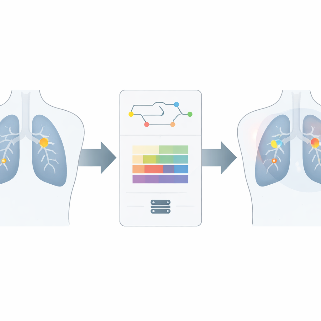
Lung cancer often begins as tiny round spots called nodules on routine chest X-rays. These specks can be easy to miss, even for experienced radiologists who review hundreds of images a day. The study behind this article describes a new artificial intelligence (AI) system that helps doctors spot these early warning signs more accurately and more quickly, using a refined version of a popular real-time vision algorithm.
The challenge of finding tiny spots
On an X-ray, lung nodules can be very small, faint, and partly hidden by ribs, blood vessels, or the heart. Radiologists must carefully search every corner of each image, a demanding task as medical imaging volumes rise worldwide. Missed nodules can delay diagnosis, while false alarms can trigger anxiety and extra tests. An ideal computer helper would reliably highlight suspicious spots without slowing workflow or requiring costly hardware.
An upgraded digital detective
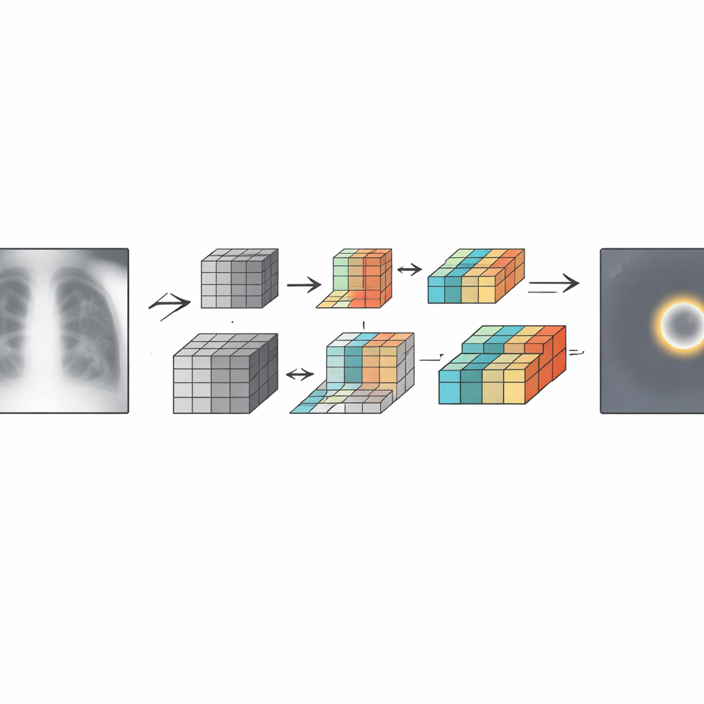
The researchers built on YOLOv12, a fast, single-pass object detection algorithm widely used in computer vision tasks ranging from traffic monitoring to industrial inspection. Standard YOLOv12 is already efficient, but it struggles with very small, low-contrast targets such as lung nodules. To address this, the team created an improved version called YOLOv12-DSV by adding three key building blocks that help the network preserve fine details, enhance tiny structures, and run leaner.
Keeping details sharp inside the model
The first building block, called space-to-depth convolution, reorganizes image information so that fine spatial details are not discarded when the image is shrunk inside the network. This is especially important for nodules that may span only a few pixels. The second, a dynamic upsampling step, reconstructs higher-resolution feature maps in a content-aware way rather than using fixed formulas, helping restore crisp edges and subtle textures around candidate nodules. The third component, a lightweight feature module, cleverly reuses and combines information so the model can focus on the most informative patterns without becoming bulky or slow.
Testing on thousands of chest X-rays
To judge whether these changes really matter, the authors trained and tested their system on two public collections of chest X-rays from the Roboflow platform, each image carefully labeled by experienced radiologists. They used cross-validation to guard against chance findings and compared their model not only with the original YOLOv12 but also with earlier YOLO versions and other leading detection methods. The improved model detected more true nodules while reducing missed cases and slightly cutting down false alarms. It also ran faster and with fewer internal parameters than the original, meaning it could be easier to deploy on standard clinical hardware.
Strengths, limits, and real-world use
The upgraded system showed the best balance between accuracy and speed among the approaches tested, suggesting it could serve as an efficient second reader for radiologists. Still, the work has limits. The datasets, though carefully annotated, may not capture the full diversity of patients, machines, and scanning protocols seen in everyday practice. The images mostly contained confirmed nodules, so the model was not trained to distinguish normal from abnormal exams on its own. The authors also note that performance on low-power devices and across different hospitals remains to be explored.
What this means for patients
In plain terms, this study shows that a thoughtfully redesigned AI engine can spot small lung nodules on X-rays more accurately while using less computing power. It does not replace the radiologist, but it could act like a tireless assistant, pointing out subtle findings that deserve a closer look. With further testing on larger and more varied datasets, tools based on this approach may help catch lung cancer earlier and make chest X-ray screening more reliable and scalable.
Citation: Mao, M., Hong, C., Zhang, Y. et al. Research on lung nodule detection in X-ray plain films based on improved YOLOv12 model. Sci Rep 16, 11667 (2026). https://doi.org/10.1038/s41598-026-47670-9
Keywords: lung nodules, chest X-ray, deep learning, object detection, medical imaging AI