Clear Sky Science · en
Machine-enhanced reconstruction of functional connectomes unravels discriminative brain sub-systems in health and disease
Why this research matters
Autism is linked to differences in how brain regions talk to each other, but standard brain scans often blur these patterns. This study shows how combining brain imaging with modern machine learning can reveal hidden communication pathways in the brain that better distinguish people with autism from neurotypical individuals, opening new doors for research on brain disorders.
Looking at brain activity as a network
The authors start from a simple idea: the brain can be viewed as a vast network, where regions are like cities and their communication links are like roads. Using functional MRI, they record how hundreds of brain regions fluctuate over time while people rest. Instead of just noting whether two regions are correlated, they organize these relationships into multilayer networks that capture activity at different frequency bands, much like separating the bass, mid, and treble channels in music. This richer picture preserves details about how brain systems coordinate across timescales.
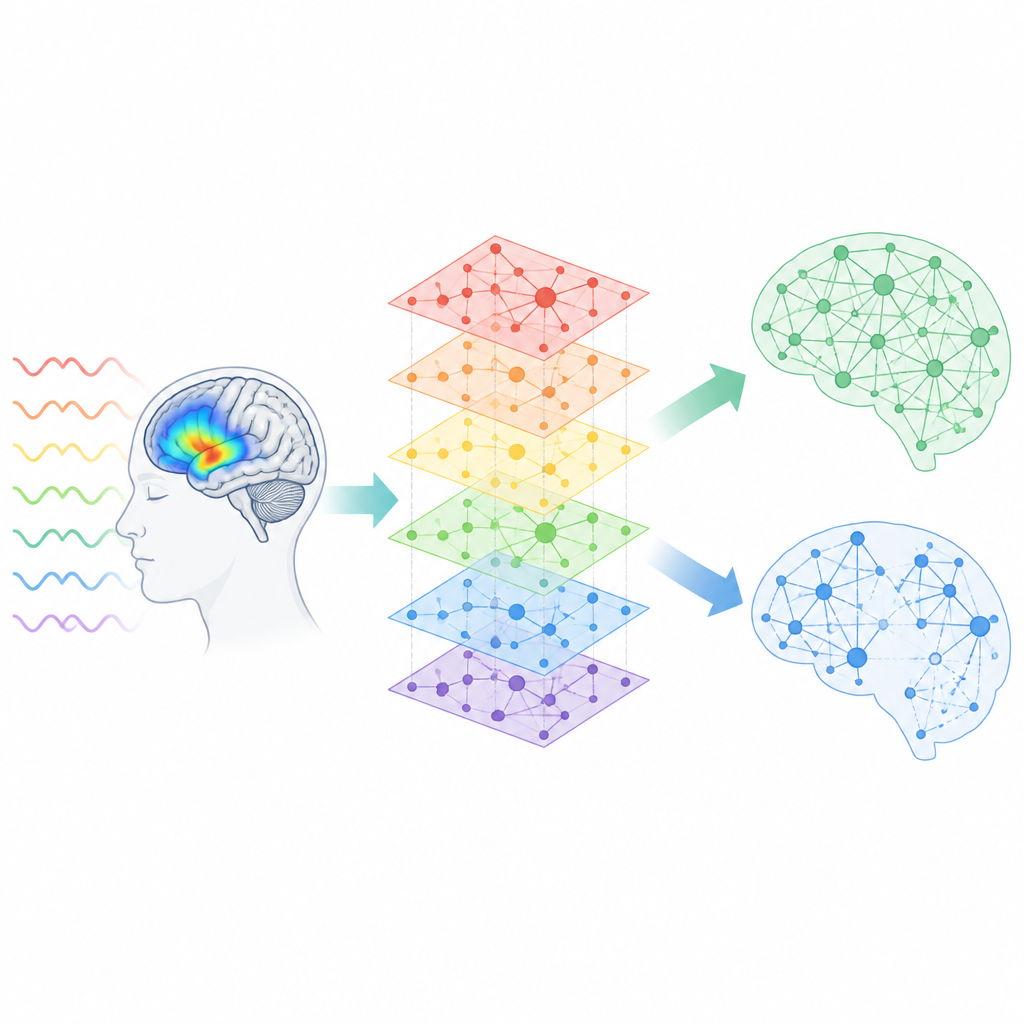
From raw scans to the most telling connections
Conventional methods prune brain networks by keeping only the strongest pairwise links, based on a fixed statistical threshold. However, this approach treats each connection in isolation and can miss meaningful patterns formed by groups of weaker links that work together. The researchers propose a different route, which they call functional pruning. They first build frequency-specific brain networks that have already been cleaned using a rigorous statistical procedure, then feed those networks into a graph-based deep learning model that learns to classify each person as autistic or neurotypical purely from the pattern of connections.
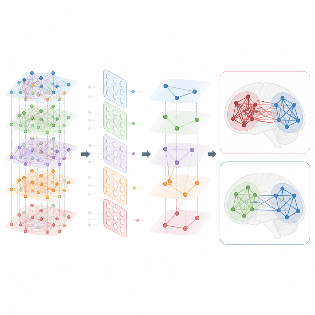
Letting the machine highlight key sub-systems
Once the model learns to distinguish the two groups, the team uses an explainable artificial intelligence tool to ask which connections mattered most for its decisions. This tool assigns an importance score to every link in the network. By gradually removing links with low scores, they discover that the model not only keeps working when over 90 percent of connections are dropped, but actually becomes more accurate at a particular pruning level. The remaining links form compact sub-systems of brain regions whose collective co-activation is especially informative about whether a person has autism or not. In other words, functional pruning favors groups of connections that work together, rather than single standout links.
What changes in autism are revealed
With these pruned networks in hand, the researchers use tools from network science to compare people with autism to neurotypical controls. They find that, after functional pruning, several network measures reliably differ between the two groups, while the original unpruned networks show no meaningful differences. In the autism group, brain sub-systems tend to have more connections on average and to form more tightly knit clusters, suggesting a pattern of hyper-connectivity. These changes are most pronounced in specific large-scale systems, including the default mode network, fronto-parietal control regions, visual areas, somatosensory regions, and subcortical structures such as the thalamus, and are especially strong in particular low-frequency bands of brain activity.
Reorganizing the brain’s communication map
Beyond simply being more connected, the brains of people with autism show a different large-scale organization. When the team examines how whole brain systems group together, they observe that networks involved in internal thought, sensory processing, and control functions cluster differently in autism. For example, certain control and salience systems, which help the brain decide what information is important, shift their relationships with other subsystems. This suggests that autism involves not only stronger connections within particular circuits but also a broader reshaping of how brain systems are arranged in a hierarchy.
What this means for understanding brain disorders
By combining functional MRI with graph-based deep learning and explainable AI, this work shows that examining collective patterns of connectivity can reveal subtle but reliable differences between autistic and neurotypical brains. Rather than relying on arbitrary cutoffs for individual links, functional pruning focuses on the sets of connections that best separate health from disease. Although this study focuses on autism, the same strategy could be applied to other brain conditions, and more generally to any complex system where we can record many activity signals but do not know the underlying wiring in detail.
Citation: Grassia, M., d’Andrea, V., Finc, K. et al. Machine-enhanced reconstruction of functional connectomes unravels discriminative brain sub-systems in health and disease. Sci Rep 16, 16173 (2026). https://doi.org/10.1038/s41598-026-47391-z
Keywords: autism, brain connectivity, functional MRI, machine learning, graph neural networks