Clear Sky Science · en
Magnetic resonance imaging diagnosis of knee injuries after skiing in adolescents under deep learning
Why knee scans matter for young skiers
Skiing is increasingly popular with teenagers, but it also brings a real risk of knee injuries that can affect both performance and long term joint health. Doctors rely on magnetic resonance imaging (MRI) to see inside the knee, yet small tears and early cartilage damage can be easy to miss when scans are read by eye. This study explores how an artificial intelligence system can help radiologists read knee MRI images from adolescent skiers more quickly and more consistently, catching subtle problems before they grow worse.
How skiing stresses young knees
Adolescent skiers face a high rate of knee problems, especially tears of the anterior cruciate ligament and meniscus, as well as cartilage damage and strains of the medial collateral ligament. These injuries can interrupt training, raise the chance of future arthritis, and even shorten sports careers. At the same time, teenagers are not just small adults. Their bones and cartilage are still developing, so injuries look different on scans and may occur in other locations than in mature athletes. Despite this, most computer tools for reading knee MRI have been trained on adult images, leaving a gap in support for young competitors.
Limits of reading scans by eye
Standard MRI diagnosis depends heavily on the skill and experience of the radiologist. Reading a full set of knee images slice by slice is slow and tiring, and tiny defects in cartilage or partial ligament tears may be overlooked. Different doctors may also judge the same scan differently, which can complicate treatment decisions. Earlier computer methods tried to help by measuring simple image features and feeding them into classic algorithms, but they struggled to capture the rich patterns needed to separate normal teenage anatomy from early injury.
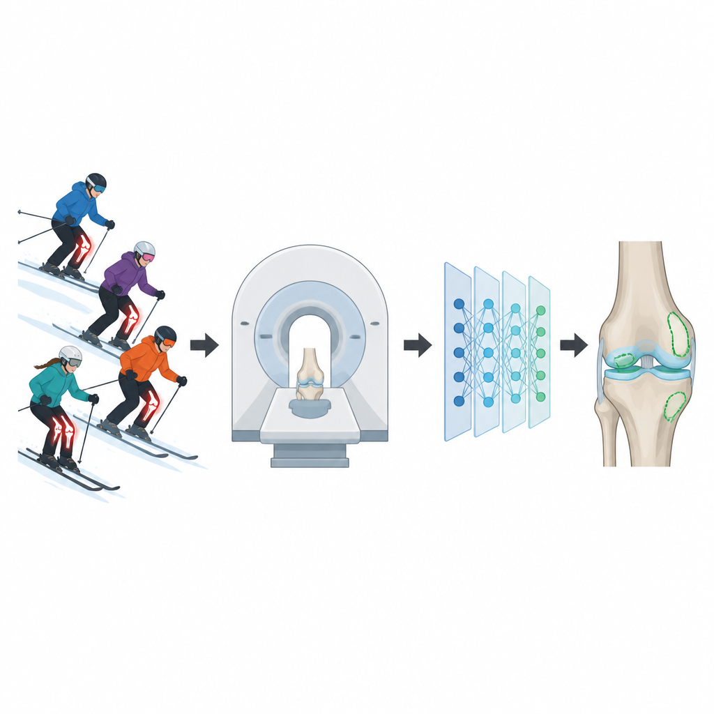
A smart model that both finds and names injuries
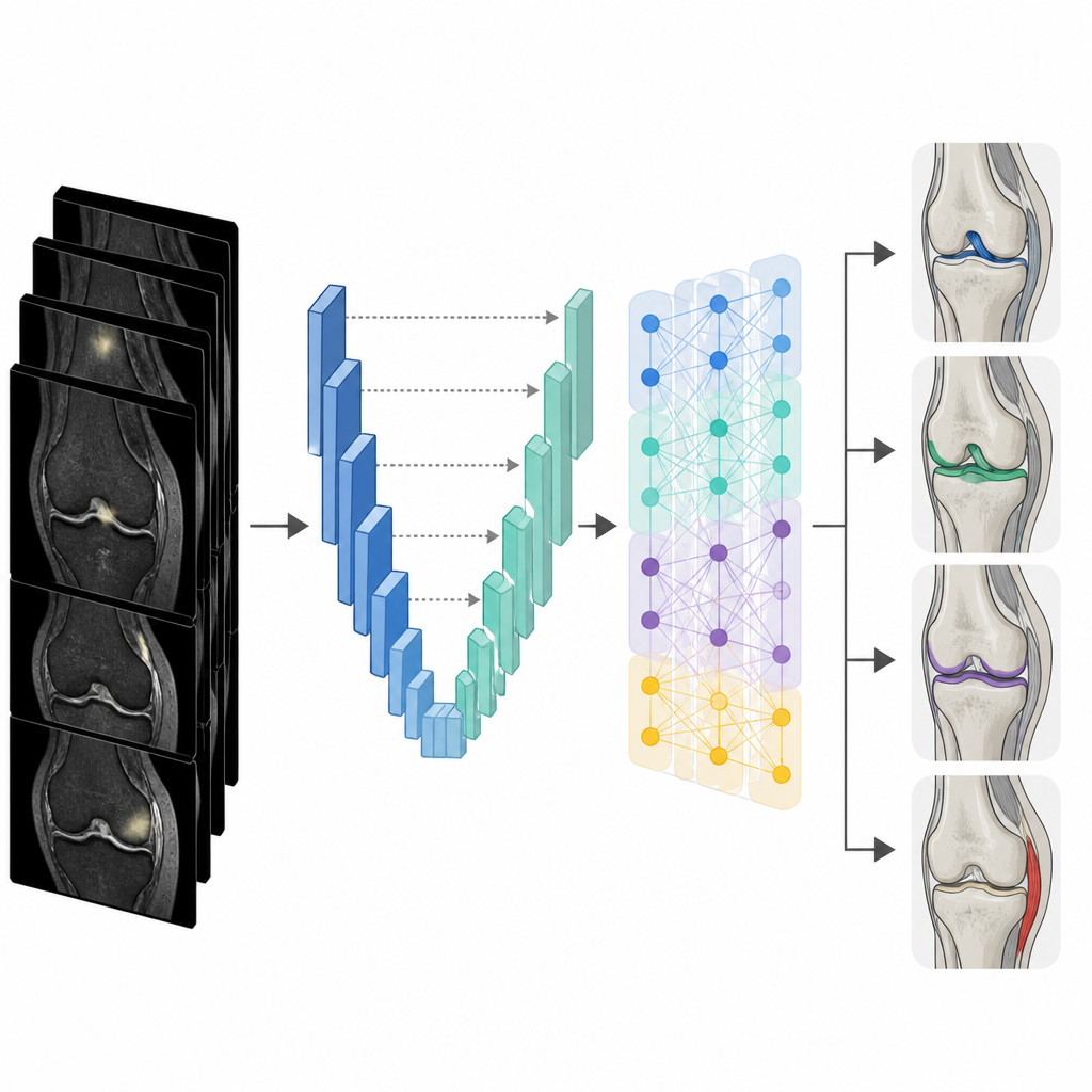
The researchers built a hybrid deep learning system that performs two tasks at once: it first outlines the exact injured regions on the MRI and then decides which types of injury are present. One part of the model, called U Net++, acts like a digital highlighter that traces four kinds of damage at the pixel level: tears of the anterior cruciate ligament, meniscus tears, cartilage injury, and medial collateral ligament strain. Its design allows it to reuse image details at many scales and receive extra guidance during training, which is especially helpful for tiny or faint lesions. The second part, based on a network called DenseNet121, takes both the original scan and the highlighted injury regions, learns patterns across them, and outputs which injuries are present in each knee.
Testing on real MRI scans from young skiers
The team trained and evaluated the system on 309 knee MRI cases from adolescent skiers aged 12 to 18. Experienced radiologists carefully drew the injury regions and agreed on final labels, providing a high quality reference for the model. The images covered several common MRI sequences and were standardized in size. During training, the system saw many slightly altered versions of each scan, such as small rotations and brightness changes, to help it cope with real world variation. The researchers compared their approach with traditional machine learning using hand crafted features, a popular deep learning network called ResNet50, and a simpler combination of separate segmentation and classification models.
How well the system performed
The hybrid model showed clear gains in both outlining and recognizing injuries. On average, its segmentation closely matched the radiologists’ drawings, especially for meniscus tears and ligament injuries, and it improved cartilage mapping compared with a standard U Net. For classification, it correctly identified injury types about nine times out of ten, with a high balance between catching true injuries and avoiding false alarms. The system outperformed both traditional feature based methods and ResNet50 across all key measures, and did so with better training efficiency than running separate models.
What this means for young athletes and clinics
In plain terms, the study suggests that a well designed AI assistant can help doctors read knee MRI scans from adolescent skiers more quickly and consistently, especially for small or easily overlooked injuries. By automatically pointing to suspicious regions and indicating likely injury types within a few seconds, such a tool could reduce missed findings, support less experienced radiologists, and speed up decisions about treatment and return to sport. The authors note that more diverse data from multiple hospitals are still needed, but their results point toward AI becoming a practical partner in protecting young knees on the slopes.
Citation: Xu, W., Li, S., Zhang, G. et al. Magnetic resonance imaging diagnosis of knee injuries after skiing in adolescents under deep learning. Sci Rep 16, 15576 (2026). https://doi.org/10.1038/s41598-026-47058-9
Keywords: knee MRI, adolescent skiing injuries, deep learning diagnosis, sports medicine imaging, ligament and cartilage tears