Clear Sky Science · en
Modelling of hybrid deep ensemble learning based skin lesion detection using FCNN denoising and inception-dilated ResNetV2
Why this matters to everyday health
Skin cancer is one of the most common cancers worldwide, yet many spots on the skin still go unnoticed until they become dangerous. This study explores how advanced computer systems can help doctors spot worrisome skin lesions earlier and more reliably by learning from thousands of images, potentially easing the burden on busy clinics and improving chances of timely treatment.
Teaching computers to read skin photos
The researchers focus on dermoscopic images, which are close-up, magnified photos that reveal fine details of moles and other skin marks. Interpreting these images is challenging even for trained specialists, especially when pictures contain hairs, shadows, or camera noise. The team aims to build a computer-aided diagnosis system that can sort different kinds of skin lesions, including melanoma, with high accuracy while handling the imperfect and uneven quality of real-world images.
Cleaning the picture before making a judgment
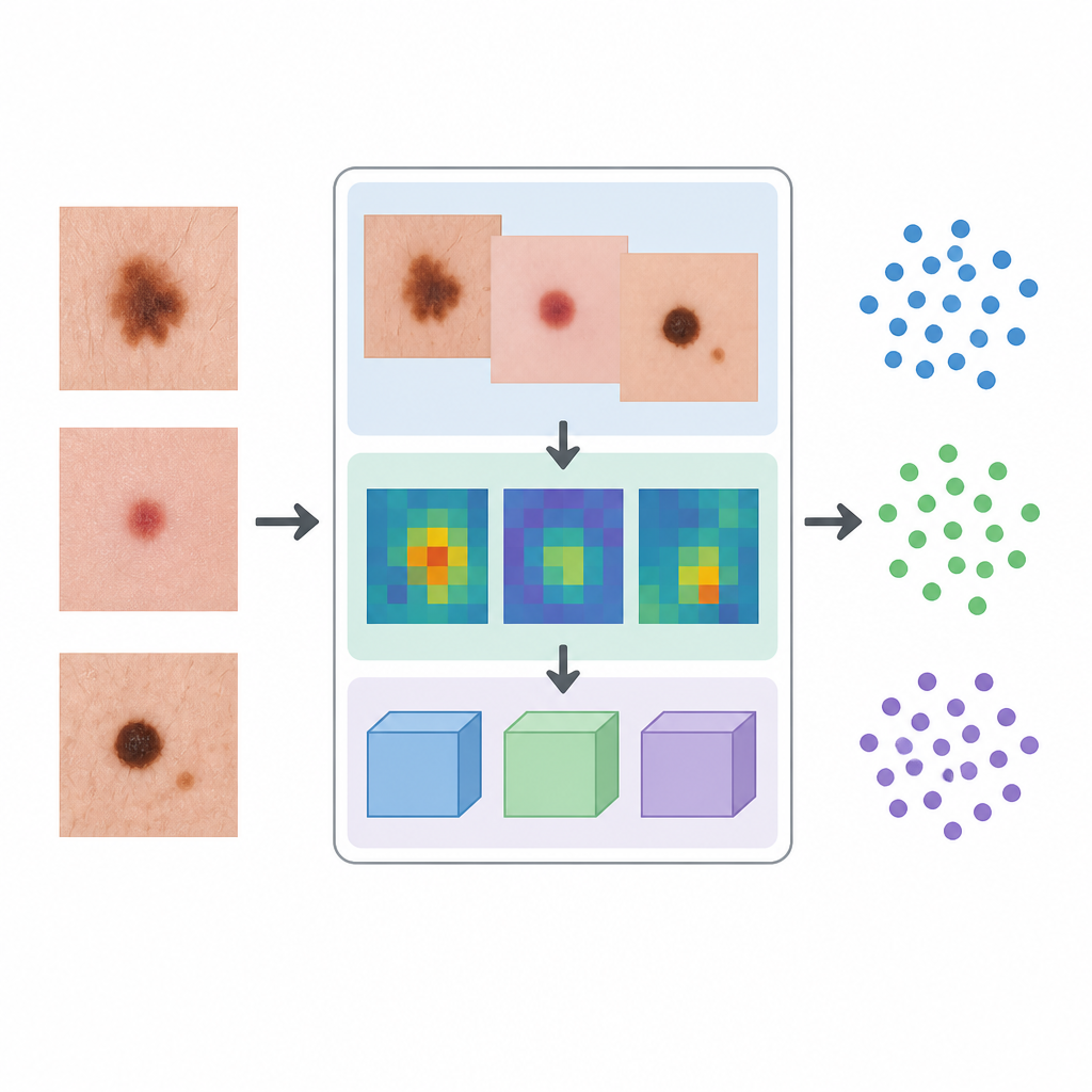
The first step in their system is to clean up each skin image using a type of neural network designed for image repair. This model learns to remove speckles, artifacts, and other visual noise while preserving the edges and textures that define a lesion’s shape and color. By training on multiple related images at once and comparing similar regions across them, the method produces a clearer version of the skin spot. This cleaner input reduces the risk that stray hairs, lighting glare, or background texture will mislead later stages of the algorithm.
Finding hidden patterns at many scales
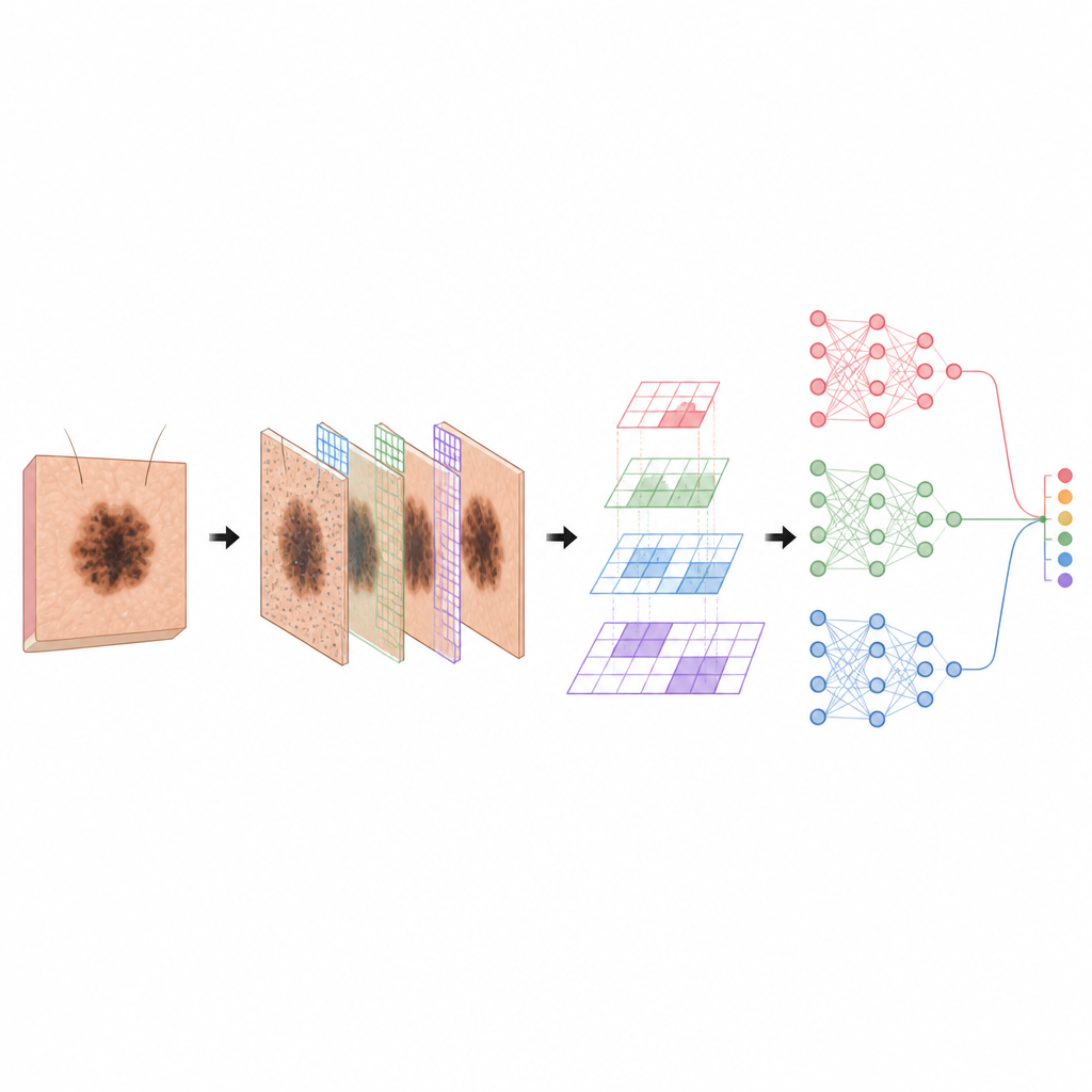
Once the images are denoised, a second deep network looks for patterns that the human eye may not easily separate. This network is built to capture both fine details, like tiny irregular borders, and broader patterns, such as overall color spread. It does so by using different filter sizes and a special layout that lets the system see both local details and wider context without exploding the amount of computation. Residual connections help information flow smoothly through many layers, so the model can build rich descriptions of each lesion without losing track of important visual cues.
Combining several "opinions" for a final call
Rather than relying on a single classifier, the authors use a trio of different deep learning models that each brings a different strength. One model is good at capturing ordered relationships in the extracted features, another learns layered statistical structure, and a third mimics spiking activity inspired by brain cells and is efficient at handling timing and pattern changes. The system blends their outputs using a weighted rule so that each model influences the final decision in proportion to how well it performs on validation data. This ensemble is trained and tested on a nine-class public skin cancer dataset that includes melanoma and several other common lesion types.
How well the system performs and what still needs work
On this dataset, the combined system correctly classifies about 99% of test images and reaches a high score for separating lesion types, outperforming a range of earlier deep learning approaches. It also runs quickly and uses less memory than several popular vision transformer models, which makes it more practical for real-world use. However, the authors note that performance drops somewhat for rare lesion categories with few examples, and that the data come from relatively similar imaging conditions and populations. They caution that generalizing to different clinics, cameras, and skin tones remains an open challenge, and that medical staff will need clear explanations before trusting automated outputs.
What this means for patients and clinicians
In simple terms, this work shows that carefully designed combinations of image cleaning, pattern discovery, and multiple deep learning models can help computers distinguish between many types of skin lesions with high accuracy on a research dataset. While such tools are not a replacement for dermatologists, they could one day serve as a smart assistant that flags suspicious spots, supports second opinions, and helps extend expert-level screening to areas with few specialists. Before that happens, larger and more diverse studies, better handling of rare cases, and closer integration with clinical practice will be needed.
Citation: Sunkavalli, J.P., Pustokhin, D., Laxmi Lydia, E. et al. Modelling of hybrid deep ensemble learning based skin lesion detection using FCNN denoising and inception-dilated ResNetV2. Sci Rep 16, 15824 (2026). https://doi.org/10.1038/s41598-026-47053-0
Keywords: skin cancer, dermoscopy, deep learning, image denoising, ensemble models