Clear Sky Science · en
MRI-based brain tumor prediction using convolutional neural network framework
Why this matters for patients and doctors
Brain tumors are often life threatening, yet spotting them early on medical scans can be difficult and time consuming. This study explores how a smart computer program can quickly read brain MRI images and flag tumors with accuracy close to that of expert radiologists, while still being light enough to run in hospitals that do not have powerful computers. The goal is to provide doctors with a fast second opinion that helps catch tumors sooner and guide treatment decisions.
How computers read brain scans
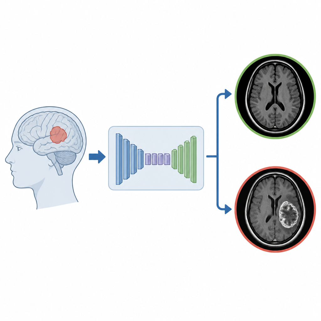
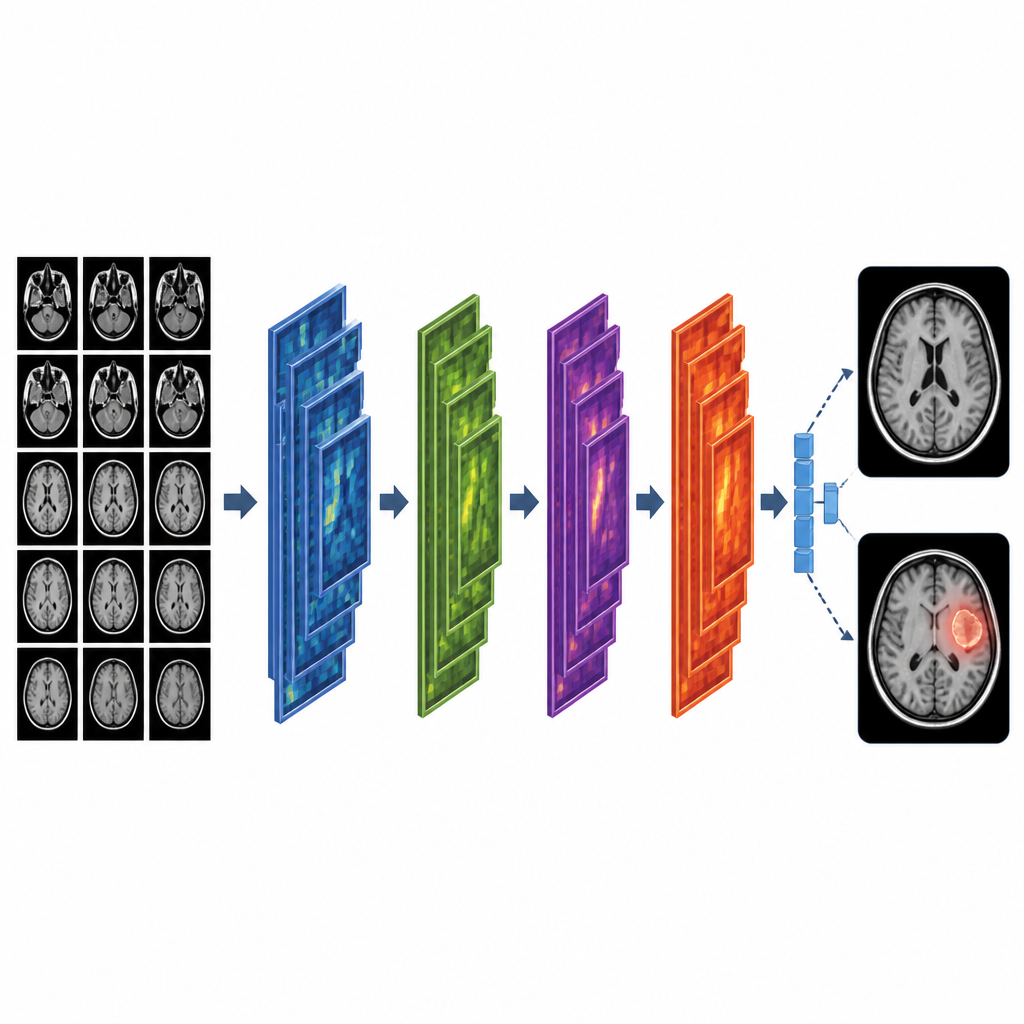
The researchers built a computer vision system based on a type of artificial intelligence called a convolutional neural network, or CNN. Instead of a human looking at each MRI slice, the network learns patterns in thousands of brain images and then uses those patterns to decide whether a new scan is healthy or shows a tumor. To train it, the team used 4600 MRI images from a public collection, labeled simply as healthy or tumor. The images came from different viewing angles, which helps the model cope with the many ways a tumor can appear in real life.
Cleaning and preparing the images
Before teaching the computer, the authors carefully prepared the MRI data so that the model would learn from clean and consistent pictures. All images were resized to the same shape and their brightness levels were scaled to a common range. Non-brain regions were removed so the network focused only on brain tissue. To prevent the system from memorizing specific scans instead of learning general rules, the team created extra training examples by flipping, rotating and slightly changing the brightness of the images. They also split the data into separate training, validation and test sets so that the final scores would reflect how the model behaves on new, unseen patients.
Designing a simple but strong model
The CNN itself was kept relatively lightweight compared with many deep learning models. Early layers detect basic structures such as edges and textures in the MRI slices, while deeper layers capture more complex shapes that are typical of tumors. The network gradually compresses this information into a small set of features and finally produces a single value that indicates whether a tumor is present. The team tuned settings such as learning rate, batch size and number of filters with a grid search, while using early stopping and automatic checkpoints to stop training as soon as the model stopped improving.
Testing how well it works
To judge whether the system would be useful in a clinic, the authors looked at a full set of performance scores, not just overall accuracy. On the held back test data, the model correctly labeled about 99 percent of both healthy and tumor scans. Sensitivity, which reflects how often true tumor cases are caught, and specificity, which measures how often healthy brains are correctly cleared, both reached 99 percent. A receiver operating characteristic curve showed an area under the curve close to one, signaling that the model rarely confuses healthy and diseased images. A confusion matrix confirmed that only a small handful of scans were misclassified out of hundreds of cases.
How it compares and what comes next
The authors compared their results with several recent AI methods for brain tumor detection. Many earlier approaches used very deep or hybrid networks that achieved high accuracy but required heavy computing power and were harder to interpret. In contrast, this study shows that a carefully designed lightweight CNN, combined with thoughtful image preprocessing and training safeguards, can match or surpass those results. The model reached 99 percent accuracy while remaining compact enough for hospitals with limited hardware and time.
What this means for real-world care
In plain terms, the study shows that a streamlined AI system can read brain MRI scans and flag tumors with very high reliability, without needing a supercomputer. While the model still needs to be tested on images from many different hospitals and scanners, it points toward practical tools that could give radiologists an extra safety net, especially where specialists are scarce. If integrated into routine workflows, such systems could help doctors spot brain tumors earlier and plan treatment more confidently, potentially improving outcomes for patients worldwide.
Citation: Reddy, Y.A., Dubey, R.S., Prakash, R.V. et al. MRI-based brain tumor prediction using convolutional neural network framework. Sci Rep 16, 15904 (2026). https://doi.org/10.1038/s41598-026-47044-1
Keywords: brain tumor, MRI, deep learning, convolutional neural network, medical imaging