Clear Sky Science · en
Assessing the reproducibility of a subject-specific finite element modelling pipeline for the human metastatic vertebrae
Why spine strength in cancer patients matters
Many people living with cancer develop tumors that spread to the spine, quietly eating away at the bones that protect the spinal cord. Doctors must decide who needs surgery or other invasive treatment to prevent painful and dangerous breaks in these backbones. This study explores whether a modern computer modeling approach based on medical scans can reliably estimate how strong a cancer damaged vertebra is, even when different people prepare the models by hand.
From medical scan to virtual backbone
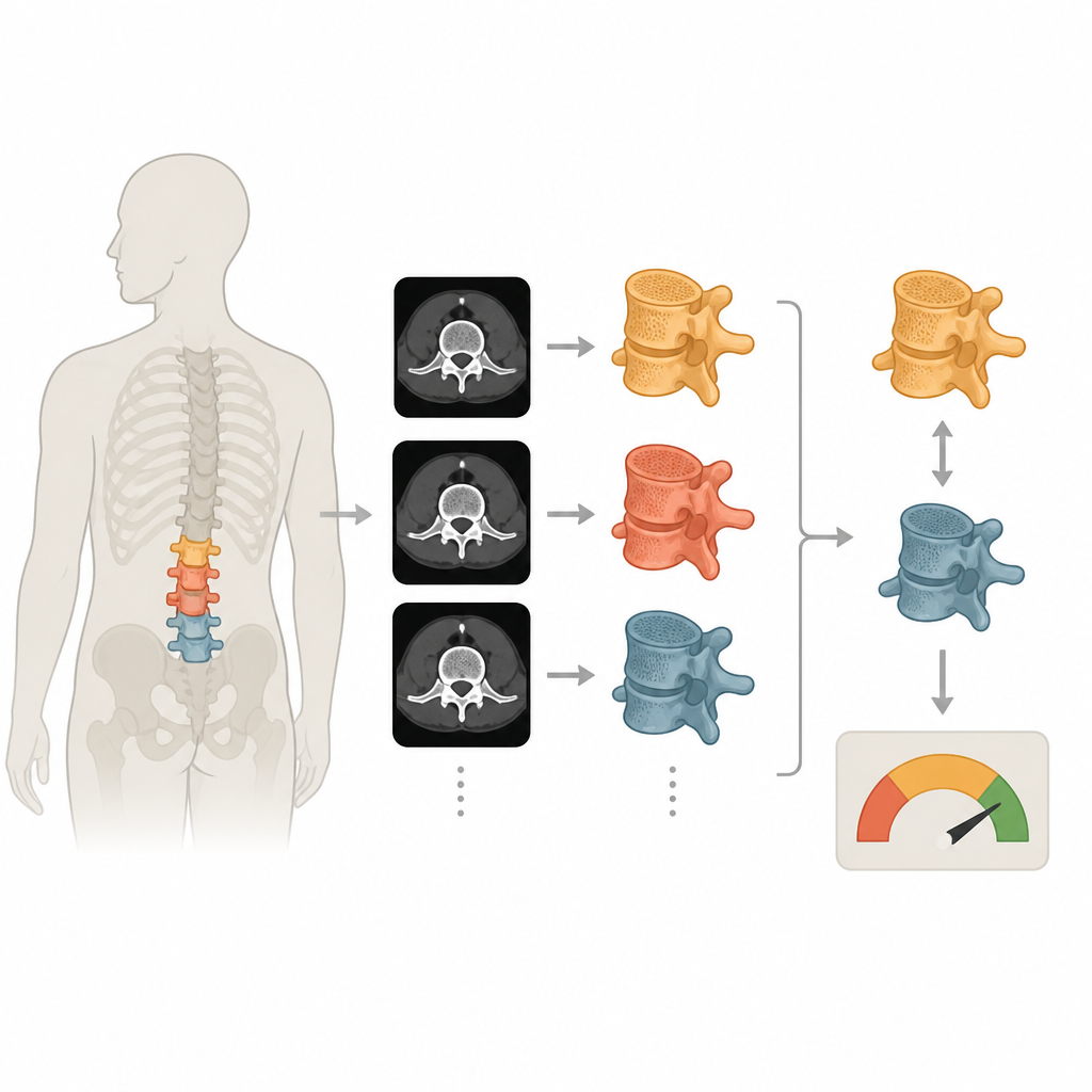
The researchers focused on a technique that turns CT scans of the lower spine into detailed three dimensional computer models of individual vertebrae. These models mimic how bone responds when it is squeezed, allowing the team to estimate how much force a vertebra can withstand before it fails. A crucial first step is tracing the outline of each vertebra on the CT images, a process called segmentation. Because cancerous lesions can blur the normal boundaries of bone, fully automatic software often struggles, so trained operators still draw these outlines manually slice by slice.
Testing consistency between human operators
To see how much these human decisions matter, the team analyzed CT scans from three patients, each with one vertebra weakened by a lytic metastasis and one nearby vertebra that appeared healthy. One experienced operator segmented every vertebra three times to test repeatability, while two additional operators each segmented them once to test differences between people. All resulting segmentations then went through the same standardized pipeline: creating a finely detailed mesh, assigning material stiffness based on local bone density, aligning the vertebra in space, and simulating a simple compression of the bone.
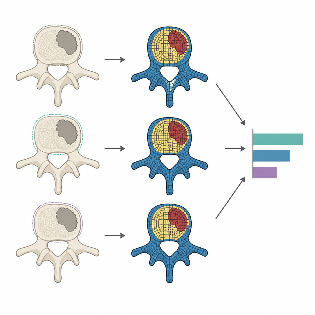
How small shape changes affect strength predictions
The scientists measured how similar the segmented shapes were using several geometry scores that compare volume, surface distance and overlap. When the same person repeated the work, the vertebra shapes were almost identical, with volume differences of about one percent and very small surface mismatches. When different people performed the segmentation, variations grew but remained modest, with volume differences around four percent and slightly larger discrepancies in some tricky regions such as bony outgrowths. Importantly, vertebrae weakened by cancer did not show a large drop in overall geometric consistency compared with their healthy neighbors.
Linking outlines to mechanical behavior
Next, the team looked at what these geometric differences meant for mechanical predictions. They examined how much force each model vertebra could carry, how stiff it appeared, and how strain was distributed throughout the bone. For the same operator, estimates of failure force and related measures varied by only about one to two percent, indicating very high stability of the pipeline. Between operators, variability roughly doubled but still stayed within a few percent for overall strength. The study also revealed that when operators consistently traced slightly different volumes, these systematic volume shifts closely tracked changes in predicted failure force, especially in vertebrae with extensive lesions where local strain estimates became less reproducible.
What this means for patient care
For a lay reader, the bottom line is that building virtual backbones from CT scans appears to be a robust way to estimate how strong a cancer affected vertebra is, as long as segmentation is done carefully and consistently. One skilled operator can reproduce their own work extremely well, and even differences between trained operators cause only modest shifts in predicted strength. This reassures clinicians and engineers that such models can support decisions about spinal stability, while also highlighting that clearer segmentation rules and future automated tools could further reduce operator related uncertainty.
Citation: Roger, R., Ghosh, R., Cai, Y. et al. Assessing the reproducibility of a subject-specific finite element modelling pipeline for the human metastatic vertebrae. Sci Rep 16, 16092 (2026). https://doi.org/10.1038/s41598-026-46900-4
Keywords: vertebral metastases, spine biomechanics, finite element modelling, image segmentation, CT based bone strength