Clear Sky Science · en
Three-dimensional reconstruction of gigapixel whole-mount histopathology specimens with RAPID
Why turning tissue slices into 3D matters
When doctors diagnose cancer, they often rely on razor thin slices of tissue viewed under a microscope. These slides reveal incredible detail, down to single cells, but they lose the original three dimensional shape of the organ. That missing depth makes it hard to compare what the pathologist sees with 3D scans such as MRI, or to measure how big and complex a tumor really is. This study introduces RAPID, a new computer method that rebuilds a full 3D picture from ordinary digital slides, using data that hospitals already collect every day.
From flat pictures to a 3D organ
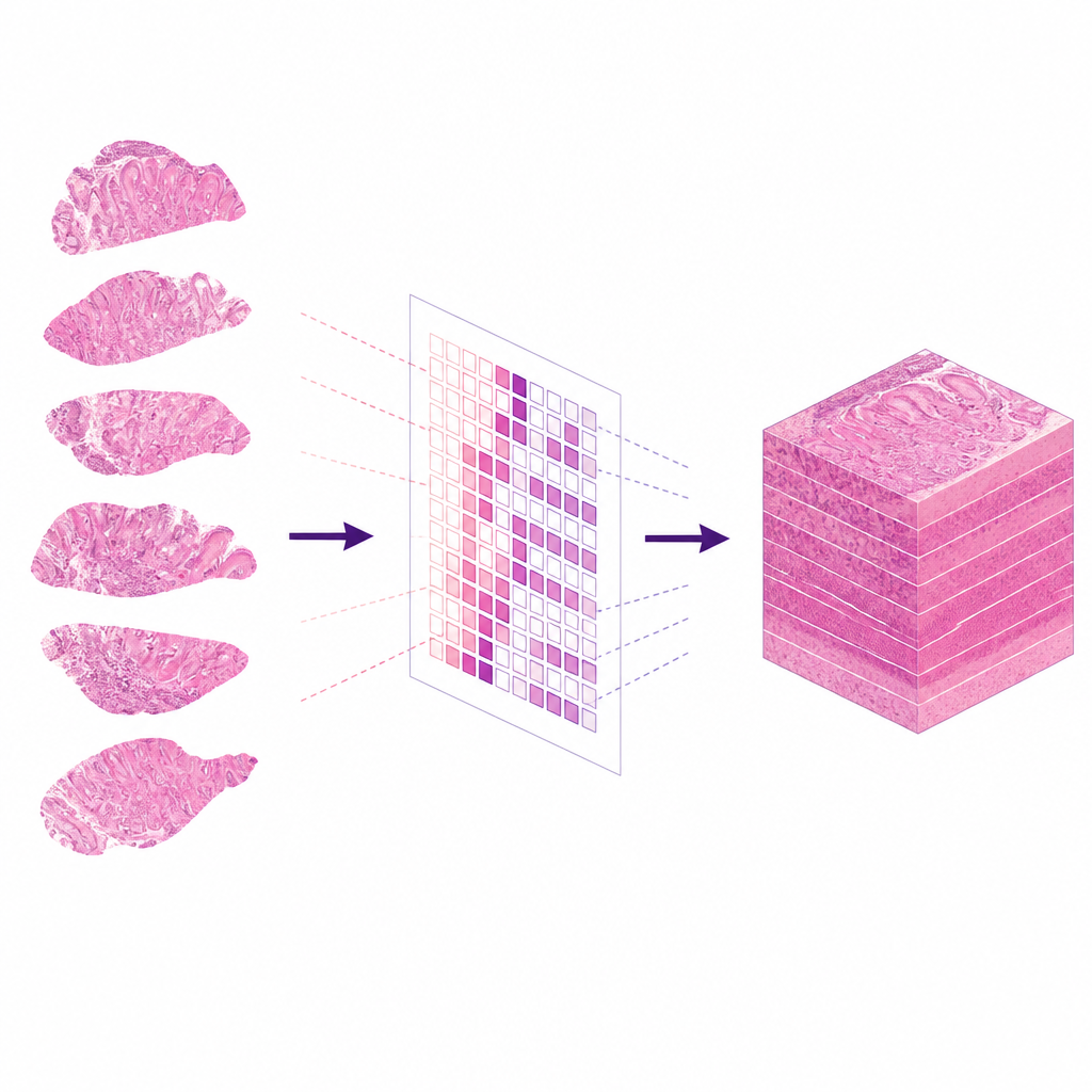
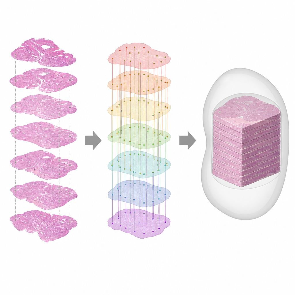
In routine care, a removed organ such as the prostate is sliced at wide intervals, stained, and scanned into extremely large images called whole slide images. Each one is a flat view of a different level in the organ, and during cutting and processing the tissue can stretch, shrink, or flip. As a result, the original 3D layout is lost and the slides no longer line up. RAPID tackles this by taking the stack of unaligned slides and computing how each one must be rotated and shifted so that, together, they approximate the original organ. The result is a virtual 3D specimen that can still be viewed at cell level detail.
Letting an AI see the big picture
A key idea in RAPID is to focus on the overall shape and large scale structures in each slide rather than on tiny matching spots. The method uses a powerful vision model, originally trained on millions of everyday photos, to extract what the authors call global features from downsampled versions of the slides. These features capture patterns like broad tissue regions and gland clusters that tend to persist even when slices are far apart. RAPID first performs a rough alignment based on the main outline of the tissue, then refines the match between neighboring slides using these global features together with finer details, while keeping the transformations anatomically realistic.
Rebuilding very large images efficiently
The original pathology images are enormous, often gigapixels in size, which would normally make such reconstructions slow and memory hungry. RAPID avoids this by doing all calculations on lower resolution copies, then scaling the resulting transformations back up to full resolution. It applies the final shifts and rotations tile by tile, streaming small blocks of pixels through memory instead of loading the entire image at once. This design allows RAPID to handle routine clinical cases on standard workstations while still producing a full resolution 3D stack that preserves cell level information.
Testing on real and challenging data
The researchers trained and validated RAPID mostly on prostate removal specimens cut at wide intervals of 4 millimeters, matching routine practice. They then tested it on additional prostate cases from other hospitals and on public datasets of mouse organs sliced much more densely. They compared RAPID with an established tool called VALIS and measured how often all slides in a case ended up with nearly correct orientation, how much the tissue shapes overlapped between neighboring levels, and how far matched structures were from one another in the final 3D result. RAPID matched VALIS on closely spaced slices but clearly outperformed it when slices were farther apart, achieving accurate reconstructions in over 90 percent of prostate cases.
Linking scans and slides in three dimensions
To show why this matters clinically, the team used RAPID to reconstruct prostates from patients who also had pre operative MRI scans. By turning the conventional slides into a 3D volume, they could visually compare the tumor seen on MRI with its true extent in the tissue. In one example, the tumor volume on the 3D reconstruction was roughly four times larger than the estimate from MRI alone, echoing known underestimation in imaging. In another, the 3D view helped relate biopsy findings to the more detailed picture from surgery. While RAPID does not yet perform full automatic matching between MRI and histology, it removes a major barrier by giving both modalities a common 3D form.
What this means for future diagnosis
RAPID shows that existing slide archives can be turned into realistic 3D models of organs without special scanners or new lab routines. For patients, this could eventually translate into better matching between imaging and pathology, more accurate estimates of tumor size, and improved tools for planning treatment. For researchers, the method opens the door to large scale studies of how diseases grow and spread through tissue in three dimensions, using data that hospitals already possess.
Citation: Schouten, D., van der Laak, J., Somford, D. et al. Three-dimensional reconstruction of gigapixel whole-mount histopathology specimens with RAPID. Sci Rep 16, 15649 (2026). https://doi.org/10.1038/s41598-026-46776-4
Keywords: 3D pathology, digital histology, prostate cancer imaging, image registration, radiology pathology correlation