Clear Sky Science · en
The necessity of cement augmentation in metastatic femur stabilization explored by finite element analysis
Why this matters for people with cancer
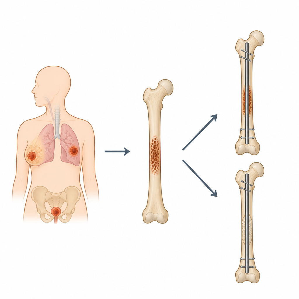
When cancer spreads to the thigh bone, a simple fall or even everyday walking can cause a serious break. Surgeons often reinforce the bone with a metal rod and sometimes add bone cement around it. This study asks a practical question that matters to patients and doctors alike: does adding cement always help, or can it sometimes make things worse?
How thigh bones are protected from breaking
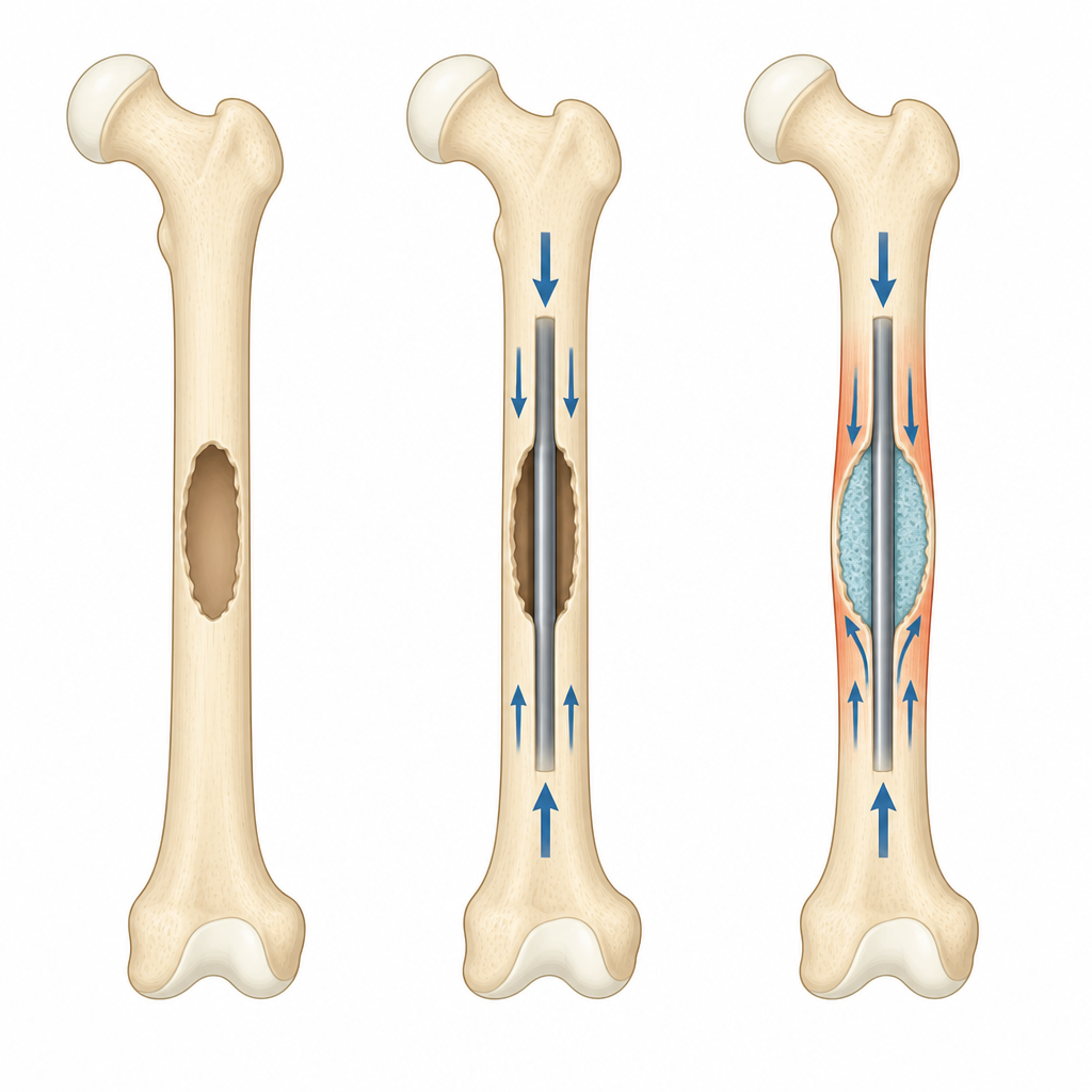
The femur is the main weight bearing bone in the leg. When cancer eats away at it, the bone can weaken enough to crack under normal daily forces. To prevent this, surgeons slide a metal rod, called an intramedullary nail, down the center of the bone to share the load and reduce pain. In some operations, they also pack the damaged area with a hard plastic material called bone cement, hoping to further support the weak spot. Yet it has not been clear whether this extra step truly improves strength in all types of lesions.
Virtual bones put to the test
Instead of testing on people or cadavers, the researchers built detailed computer models of a human femur. They created three versions of cancer damage at the middle of the bone, measuring 1, 3, and 5 centimeters in length. For each damage size, they simulated four situations: a healthy bone, a bone weakened by cancer, a bone reinforced with a nail alone, and a bone reinforced with both nail and cement. They then applied forces similar to those experienced during walking and measured how much the bone bent, how much the damaged region moved, and how much stress built up in the bone and in the metal implant.
What happens in small versus large weak spots
The nail alone clearly stabilized the bone for the medium and large defects. In the 3 and 5 centimeter lesions, it cut overall bending by about one third to two fifths compared with cancer damaged bone without surgery. Adding cement in these larger defects slightly reduced tiny movements at the cancer site and modestly lowered stress in the metal rod for the largest lesion. In contrast, for the smallest 1 centimeter lesion, filling the area with cement actually increased how much the bone and implant bent and raised the stress carried by the rod. Across all sizes, cement tended to shift more stress into nearby healthy bone because it is stiffer than natural tissue.
Hidden trade offs in stress and motion
The simulations showed that both the nail alone and the nail plus cement restored the ability of the femur to carry load, but in different ways. With cement, stress around the defect rose further, especially in smaller lesions, suggesting that nearby bone may be overworked. At the same time, micromotion at the cancer site only improved in the larger defects, while it worsened in the smallest one. The metal implant itself also felt higher stress and moved more when cement was added in small and medium lesions, which could affect how long the hardware lasts.
What this means for treatment choices
For patients with small or structurally stable spots of cancer in the femur, the results suggest that a nail alone can provide enough mechanical support without the extra step of cement. In these cases, cement may even make the mechanical environment less favorable by overloading nearby bone and the implant. For larger or more unstable defects, cement can offer added local stability, though the benefit is modest. The study supports a tailored, case by case approach, where surgeons weigh lesion size and bone quality before deciding whether to use cement rather than applying it by default.
Citation: Suh, I.W., Park, C.H. & Moon, Y.J. The necessity of cement augmentation in metastatic femur stabilization explored by finite element analysis. Sci Rep 16, 15378 (2026). https://doi.org/10.1038/s41598-026-46607-6
Keywords: femur metastasis, bone cement, intramedullary nailing, finite element analysis, pathologic fracture