Clear Sky Science · en
Changes in mRNA expression in the myocardium of rats with ventilator-induced myocardial injury
Life support and the hidden strain on the heart
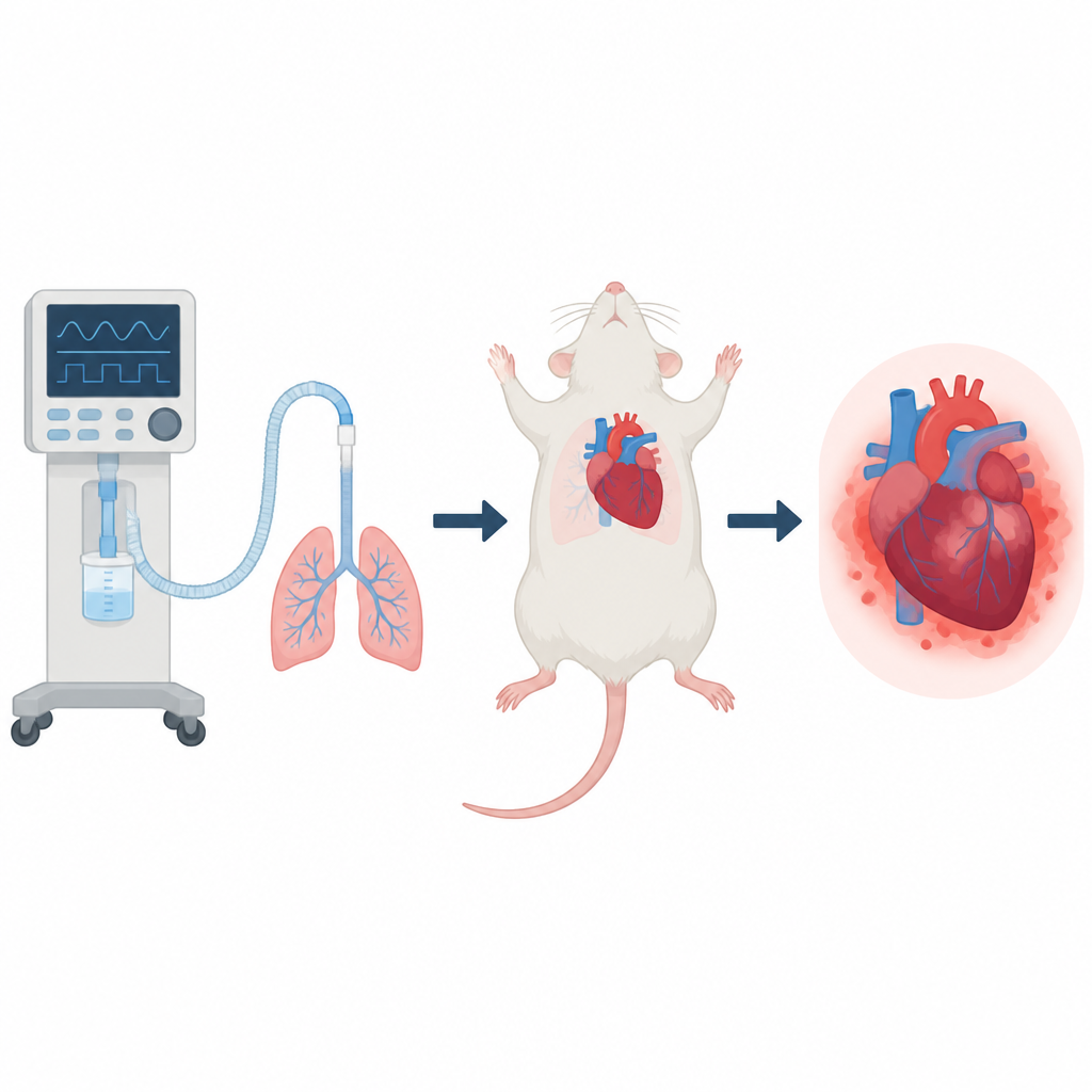
Mechanical ventilators are vital machines that help patients breathe when their lungs fail, such as during severe infections or in intensive care. But long hours on a ventilator can quietly harm more than the lungs. Doctors have noticed that some patients develop heart problems while receiving breathing support, yet the underlying reasons remain unclear. This study uses a rat model to peer inside heart cells and track how their genetic activity changes during prolonged ventilation, offering clues to why life saving support can sometimes strain the heart.
How the study was set up
Researchers worked with healthy young rats and divided them into three groups. One group underwent only the surgical preparation, serving as a baseline. The second group received six hours of mechanical ventilation, and the third received twelve hours, using settings similar to those used in human critical care. After the ventilation period, the animals were humanely euthanized and their heart tissue was examined under the microscope and with powerful genetic tools that measure which genes are switched on or off inside cells.
What the heart tissue looked like
Microscope images told a clear story. Compared with hearts from non ventilated rats, hearts from ventilated rats showed obvious damage. After six hours, the heart muscle fibers were disorganized, and there were patches of bleeding, congestion, and invading immune cells. After twelve hours, the changes were more severe: heart cells had ragged borders, the tissue was heavily congested with blood, and inflammatory cells crowded the spaces between fibers. These structural changes confirmed that mechanical ventilation alone, even in otherwise healthy animals, can injure the heart muscle and that the injury worsens with time.
Gene activity shifts toward inflammation and stress
The team then analyzed messenger RNA, the molecules that carry genetic instructions from DNA to make proteins. By comparing ventilated and non ventilated animals, they identified hundreds of genes whose activity changed, including many that were shared between the six and twelve hour groups. Many of the most strongly altered genes were involved in cell signaling, protein interactions, and binding to metal ions. A key finding was that several genes tied to inflammation and oxidative stress were turned up, while some genes that normally help protect cells were turned down. This pattern suggests that prolonged ventilation pushes heart cells into a state of heightened immune activity and chemical stress.
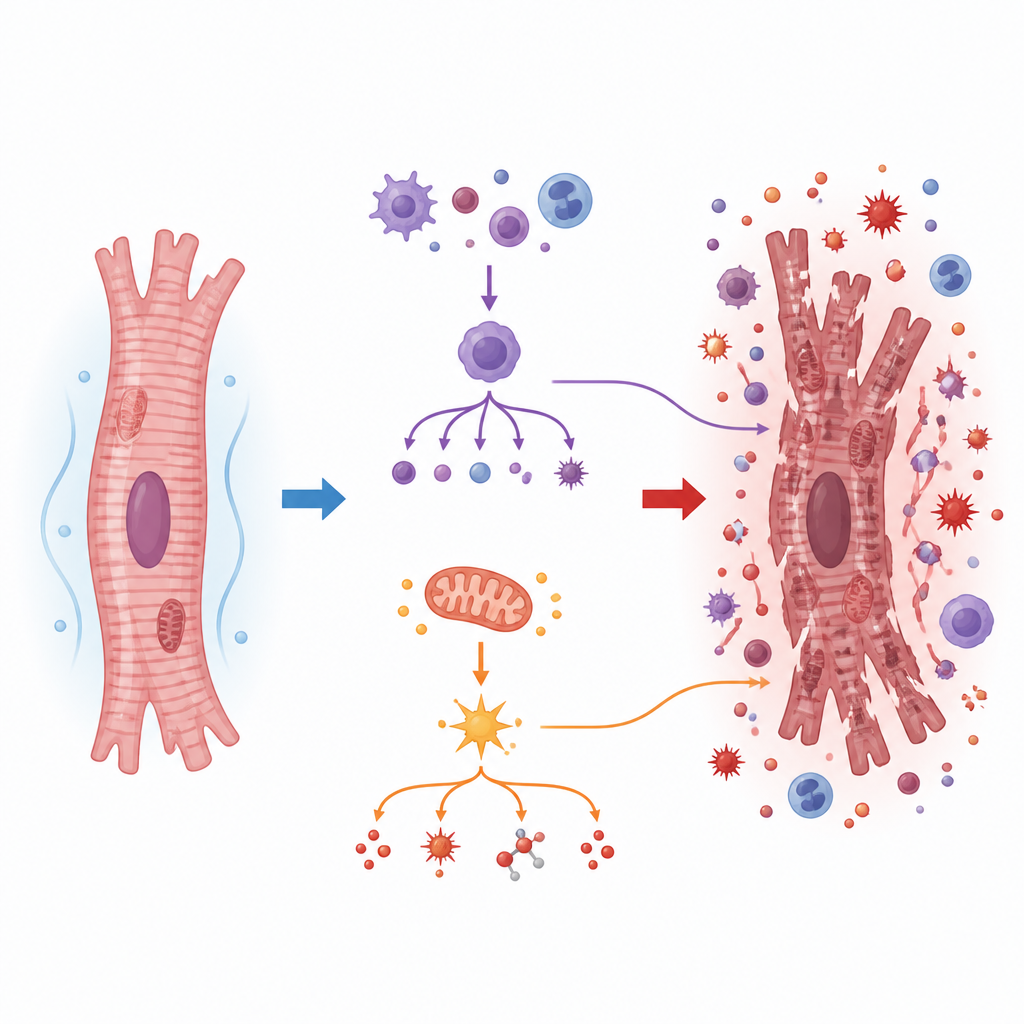
Key players and pathways inside heart cells
Among the most notable genes were C4B, TXNIP, and JUND. C4B is part of the body’s complement system, which can fuel inflammation, and its levels rose in ventilated hearts. TXNIP, which promotes the buildup of harmful reactive oxygen molecules and can trigger a powerful inflammatory complex inside cells, was also increased. In contrast, JUND, a gene that usually helps limit oxidative damage, was reduced. Together, these shifts point to a tilt toward destructive inflammation and oxidative stress in ventilated hearts. When the researchers grouped the altered genes into known biological pathways, one signaling route, called the MAPK pathway, stood out as repeatedly enriched, linking many of these genes into a common stress response network.
What this means for patients
To confirm their genetic data, the authors used a separate technique to remeasure several key genes and found that most followed the same trends, supporting the reliability of the results. While this work was done in rats and does not directly translate into treatment advice, it shows that mechanical ventilation can rewire heart cell gene activity in ways that favor inflammation and oxidative damage, and that these changes intensify with longer support. Understanding which genes and pathways are involved, especially those linked to complement activation, oxidative stress, and MAPK signaling, may help scientists design safer ventilation strategies and explore new drug targets to protect the heart during critical illness.
Citation: Liao, S., Zhao, D., Zeng, L. et al. Changes in mRNA expression in the myocardium of rats with ventilator-induced myocardial injury. Sci Rep 16, 15937 (2026). https://doi.org/10.1038/s41598-026-46579-7
Keywords: mechanical ventilation, ventilator-induced myocardial injury, heart inflammation, RNA sequencing, MAPK pathway