Clear Sky Science · en
Generative cerebral vasculature visualization using spatial transcriptomic data
Why brain blood vessels matter
The brain depends on a dense web of blood vessels that deliver oxygen and nutrients every second. When this network falters, thinking, movement and memory can quickly suffer. Yet mapping these tiny vessels across the whole brain is extremely hard. In this study, researchers use a form of artificial intelligence to turn molecular data from mouse brains into detailed virtual pictures of the brain’s blood vessel system, offering a new way to explore how healthy blood flow supports brain function.
A new way to see the brain’s plumbing
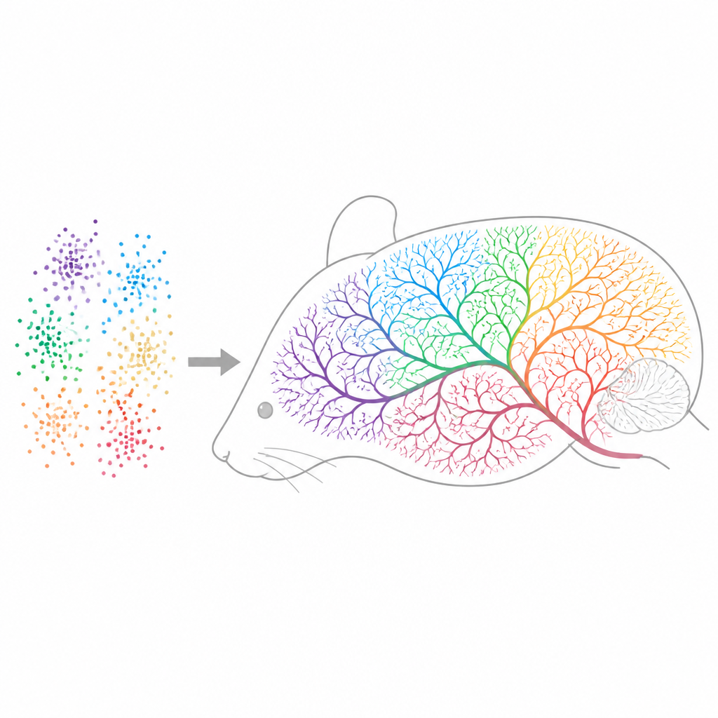
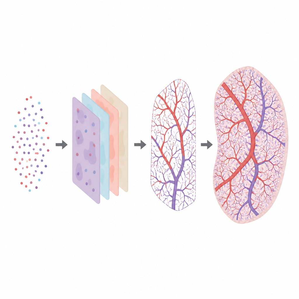
Instead of starting with traditional microscope images, the team works from spatial transcriptomics, a technique that records which genes are active and where in thin brain slices. They build on a generative model called Tera-MIND, originally developed to simulate mouse brain structure from gene activity patterns. Here, they repurpose it to focus on blood vessels. The model learns how the activity of certain vessel-related genes tends to appear in space, then uses that information to “imagine” realistic pictures of brain tissue in which vascular patterns emerge from the gene data alone.
Turning gene signals into vessel maps
The researchers concentrate on two key genes that mark different parts of the vascular system. One gene, Cldn5, is active in the thin cells that seal the blood–brain barrier in small vessels. The other, Acta2, is active in muscle cells that wrap around larger arteries. By feeding Tera-MIND the spatial activity of these genes across many sections of three mouse brains, the model produces images that show capillary-rich regions and major arterial territories where biology predicts they should be. It also calculates an internal “attention map” that highlights places where both genes are active together, which helps the model connect scattered signals into continuous, vessel-like tracks.
Testing the virtual vessels
To check whether these virtual vessels make sense, the team compares the model’s predictions with known maps of mouse brain vasculature and with patterns seen in the original imaging data. They find that predicted Cldn5 signals follow long, slender paths typical of capillaries, while Acta2 signals line up with cross-sections of larger arteries on the brain’s surface and deeper inside. Across 50 brain slices, the model’s attention maps for Acta2 show a strong correlation with where this gene is actually expressed, and regions where Cldn5 and Acta2 appear together improve predictive accuracy. This suggests the model uses the combined gene activity to infer how different vessel types are arranged and connected.
Strengths, limits and future uses
The approach offers a fast, cost-effective and animal-sparing way to explore vascular structure without new staining or complex microscopy. By reusing existing spatial gene datasets, it can generate three-dimensional views of how vascular territories span different brain regions and how gene activity relates to blood vessel organization. However, the underlying data come from brain slices separated by sizable gaps, and the gene signals are sparse. As a result, the virtual vessels should be seen as probabilistic outlines of vascular territories rather than exact reconstructions of every capillary branch. The model cannot recover fine-grained topology where gene markers are missing or rarely sampled.
What this means for brain health research
The study shows that generative AI can turn molecular snapshots into interpretable, large-scale pictures of the brain’s blood supply. By focusing on a small set of well-chosen vascular genes, the model recovers realistic patterns of arteries and capillaries across whole mouse brains. In the future, adding more vascular markers and combining this method with imaging and genomic data may help scientists probe how blood flow changes in disease models, such as those with blood–brain barrier leaks or vessel damage. For now, the work establishes a powerful in silico tool for exploring how the brain’s plumbing is organized, using information already hidden in existing gene maps.
Citation: Berg, I., Wu, J. & Koelzer, V.H. Generative cerebral vasculature visualization using spatial transcriptomic data. Sci Rep 16, 15540 (2026). https://doi.org/10.1038/s41598-026-46455-4
Keywords: cerebral vasculature, spatial transcriptomics, generative AI, mouse brain, blood brain barrier