Clear Sky Science · en
Ex vivo model for assessing fetal membrane integrity and therapeutic strategies
Why protecting the baby’s “water sac” matters
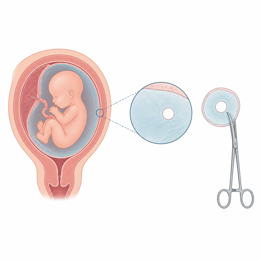
Before birth, every baby grows inside a thin but tough “water sac” made of fetal membranes that hold the amniotic fluid and form a barrier between mother and child. When this sac breaks too early, it can trigger preterm birth, which affects millions of families worldwide. Modern fetal surgeries, which can save or improve babies’ lives, often require making small holes in these membranes and can unintentionally increase the risk that the sac will later give way. This study introduces a new lab model that keeps pieces of human fetal membrane alive and functioning for weeks, so scientists can better understand how the sac weakens, how it might repair itself, and how new sealing materials or devices could help prevent dangerous early ruptures.
A closer look at the baby’s protective sac
The fetal membranes are made of two closely joined layers that together act like a flexible raincoat and security gate. The inner layer, called the amnion, sits next to the amniotic fluid and carries much of the mechanical load, while the outer layer faces the womb and helps with immune protection. Many different cell types and a rich web of supporting fibers and gel-like material give the membranes their strength and barrier function. When doctors perform procedures inside the womb, they must pierce these layers, and current sealing materials such as collagen sponges or fibrin “glues” often fail to provide stable, long-lasting closure. Animal models used to test new solutions do not fully mimic the unique structure and healing behavior of human membranes, and simple cell cultures cannot capture the complex interactions between cells, fibers, and mechanical forces.
Building a long lasting lab model of human membranes
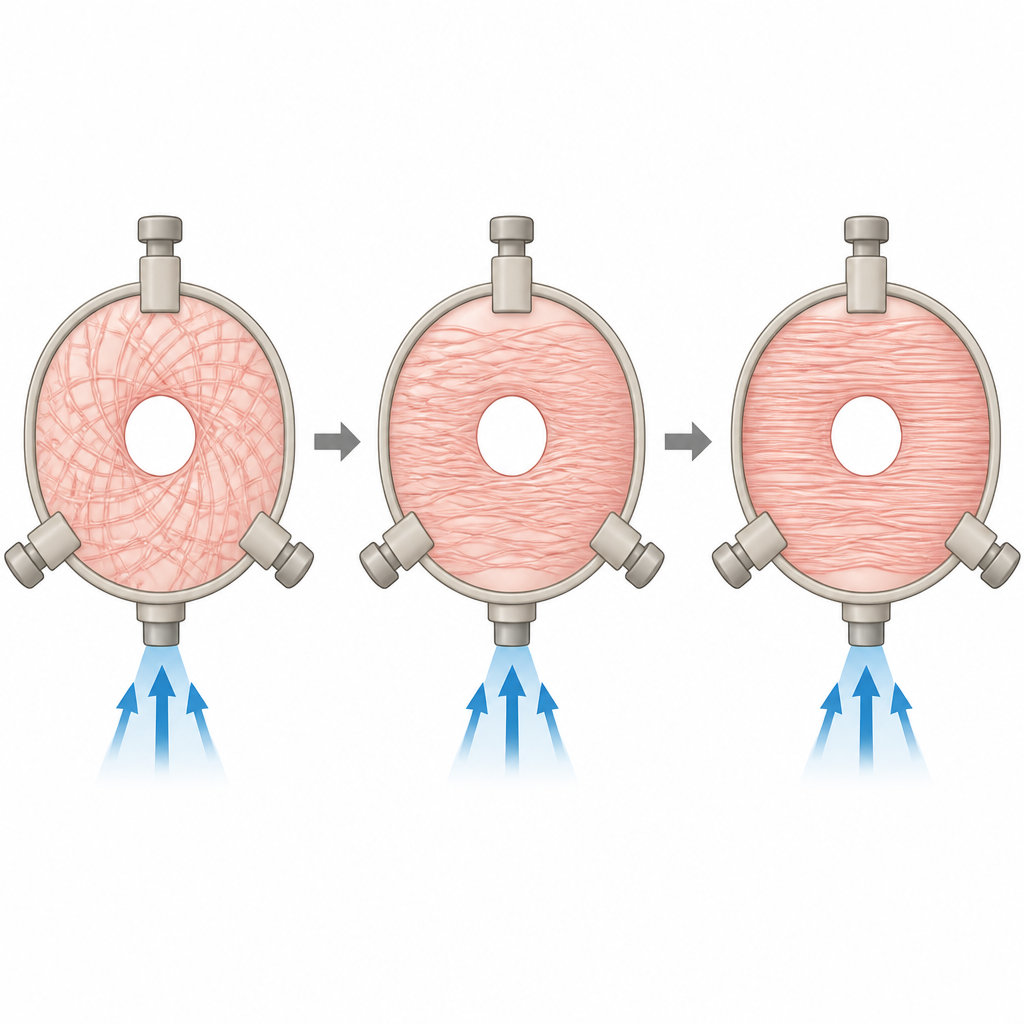
The researchers developed a 3D printed device that clamps a small round piece of full thickness human fetal membrane, preserving both inner and outer layers. The device holds the tissue under gentle tension and can be placed on a culture platform that bathes both sides in nutrient rich fluid. It is modular, allowing different media on each side, mechanical testing, and the creation of standardized holes similar in size to those made during fetoscopic surgery. Using this setup, the team kept human membranes alive for up to 21 days, far longer than most earlier systems. They tracked DNA and energy levels, sugar use, and waste production in the surrounding fluid to see how the tissue adapted over time.
What the model reveals about life and strength in the membrane
Over three weeks, the membranes largely maintained their structure, with living cells still present in the supporting layers and much of the surface lining intact, though some small gaps appeared by day 21. DNA content stayed stable, while the main energy signal, ATP, slowly declined, suggesting a gradual shift in how cells were working rather than sudden death. The barrier that keeps substances from freely crossing the membrane was mostly preserved, with only donor to donor variation. Measurements of sugar uptake and lactate release showed that metabolism first spiked as the tissue adapted to its new environment, then settled into a steady pattern. When the team used a custom pressure chamber to push on the clamped tissue from below, they found that membranes kept their rupture strength after two weeks in culture and were naturally strongest near the placenta, becoming weaker farther away.
Watching damage and repair like a slow motion movie
To mimic surgical injury, the scientists punched 3 millimeter holes into the clamped membranes, matching the size of typical access ports used in fetal surgery. Using specialized imaging, they observed how collagen fibers around these holes changed over days. The fibers gradually became more aligned around the defect, especially between days 3 and 7, a pattern also seen in samples from operated pregnancies and thought to influence whether a tear heals or stays weak. Cells near the edge of the hole also changed their shape and markers, hinting at active remodeling. When the team tested a common fibrin based glue on these larger defects, the material had dissolved completely after two weeks, underscoring why more durable sealing strategies are needed.
What this means for future pregnancies and treatments
This new ex vivo model offers a practical human based “test bed” for studying how fetal membranes respond to injury and for trying out new plugs, patches, and healing signals in conditions that closely resemble real pregnancies. While not perfect, it preserves membrane structure, barrier function, and mechanical strength long enough to explore how defects evolve and how candidate treatments behave over time. By reducing reliance on animal experiments and making it easier for many labs to run realistic tests with inexpensive 3D printed parts, this toolbox could help move safer fetal surgery and better prevention of early membrane rupture a step closer to everyday care.
Citation: Moser, L., Tschan, B., Gegenschatz-Schmid, K. et al. Ex vivo model for assessing fetal membrane integrity and therapeutic strategies. Sci Rep 16, 15395 (2026). https://doi.org/10.1038/s41598-026-46366-4
Keywords: fetal membranes, preterm birth, fetal surgery, ex vivo model, biomaterial sealing