Clear Sky Science · en
ResTransUNet: a dual-encoder hybrid network for automated liver segmentation in CT scans
Why this matters for patient care
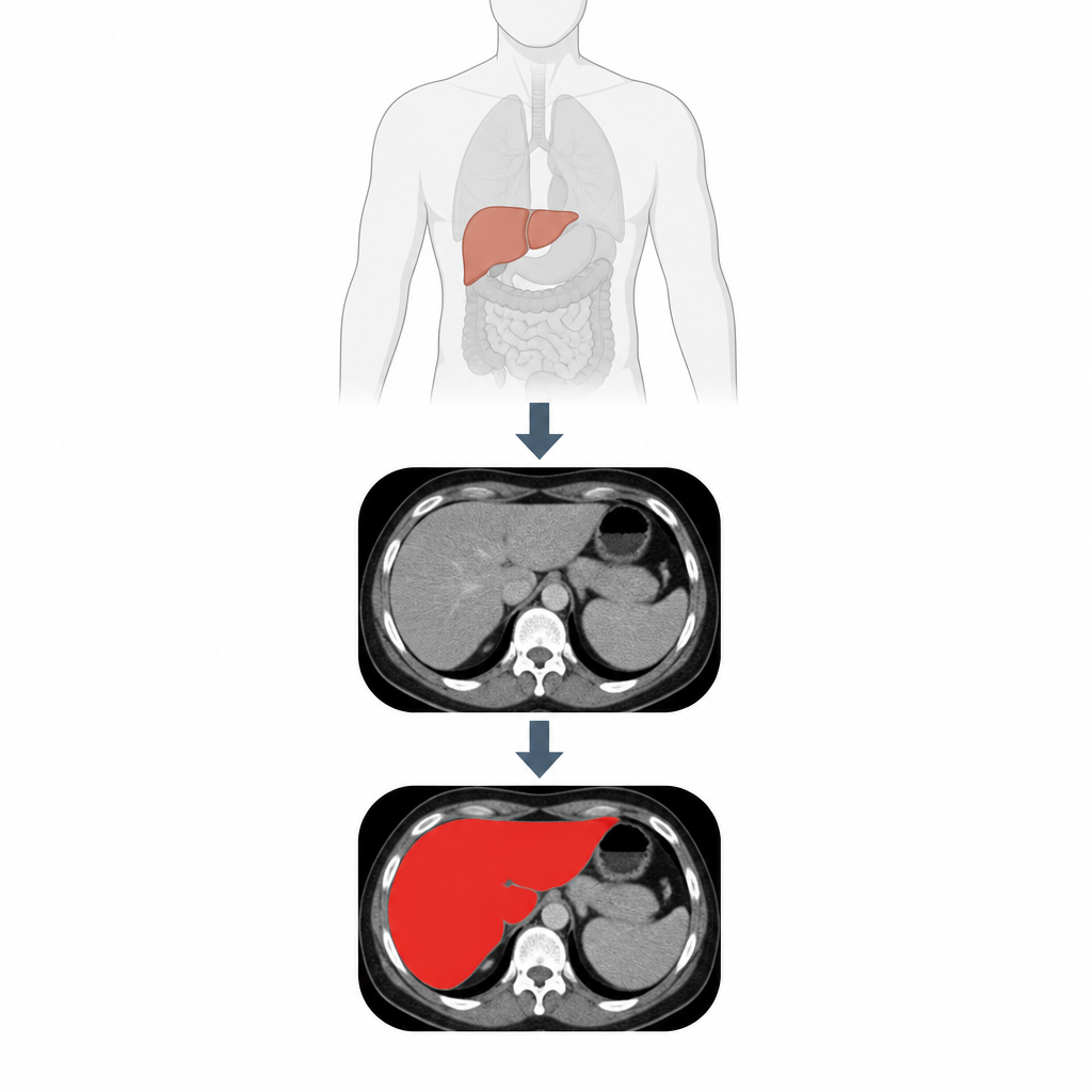
Doctors rely on CT scans to see the liver clearly when planning cancer treatment or surgery, but today many hospitals still ask experts to trace the organ by hand on hundreds of images. This is slow, tiring work that can vary from one person to another. The research described here presents a computer program that can outline the liver automatically and very accurately, which could help radiologists work faster and make liver care more consistent.
Turning raw scans into clear organ outlines
The study tackles a very practical problem: how to teach a computer to find the liver in noisy, low contrast CT images where organ borders are often fuzzy or broken. Traditional image tricks like thresholding or region growing struggle when the liver looks similar to nearby tissues. Earlier machine learning systems needed hand-crafted rules and features, which limited how well they could adapt to new patients. More recent deep learning models, especially those based on U shaped networks, improved performance by learning features directly from data, but they still miss some of the wider context in the image, which is crucial when edges are unclear.
A two track brain for seeing detail and context
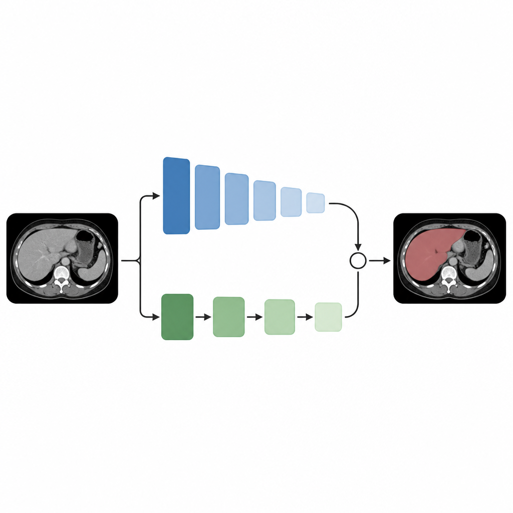
To overcome these issues, the authors introduce a model called ResTransUNet that mixes two different ways of processing images. One track is a classic convolutional pathway, inspired by the widely used U Net design, which is very good at capturing fine local details and precise shapes. The other track is built from transformer blocks, a newer family of models that excel at seeing long range relationships across an image, such as how distant parts of the liver relate to each other. The key idea is to let these two tracks run side by side and talk to each other throughout the network, so that every stage of processing blends sharp local edges with broad contextual understanding.
How the model learns what to focus on
Inside the convolutional track, the network uses residual links and channel wise attention modules to keep useful information flowing and to emphasize the most informative patterns. A special component called the Feature Enhancement Unit serves as a bridge between the transformer and convolutional tracks. At several depths in the network, it takes the global view from the transformer and the local features from the convolutions, combines them, and then learns how strongly each channel should contribute. In addition, a multi scale block looks at the image with several virtual zoom levels at once, helping the model cope with livers of different sizes and shapes and with regions that are broken into small pieces.
Testing on many types of scans
The researchers trained and tested ResTransUNet mainly on a large public liver CT collection used in an international challenge, then checked how well it transferred to three other well known datasets. They measured how much the computer drawn liver overlapped with expert outlines, how often it included too much tissue, and how much volume error remained. Across all these tests, the new model consistently scored higher than eight strong competing methods, including both classic U Net variants and other systems that already use transformers. It showed particular strength on difficult cases with small or fragmented liver regions and on scans where the liver boundary is hard to see.
From the lab to the reading room
For a non specialist, the bottom line is that this work delivers an automatic tool that can outline the liver on CT images with accuracy close to expert humans, while also working reliably across different datasets and organs. By blending a detail oriented pathway with a context aware pathway, ResTransUNet reduces missed regions and false alarms. Although the authors note that practical deployment will still require careful integration with hospital systems and testing on a wider range of scanners and patient groups, the approach shows how smart combinations of modern deep learning ideas can turn complex medical images into clear, trustworthy maps for diagnosis and treatment planning.
Citation: Wang, Y. ResTransUNet: a dual-encoder hybrid network for automated liver segmentation in CT scans. Sci Rep 16, 15366 (2026). https://doi.org/10.1038/s41598-026-46342-y
Keywords: liver segmentation, CT scans, deep learning, transformer network, medical imaging