Clear Sky Science · en
Three-dimensional ex-vivo visualization of normal and inflamed small intestine and colonic tissue using optical coherence tomography
Seeing Gut Inflammation in Three Dimensions
Inflammatory bowel diseases such as Crohn’s disease and ulcerative colitis affect millions of people and often require repeat procedures to track how the gut is doing over time. Today’s tools either show only the surface of the intestine during a scope exam or require removing tiny pieces of tissue for lab study. This research explores a light based imaging method that can view gut tissue in three dimensions without cutting it into thin slices, with the long term goal of helping doctors tell normal, inflamed, and scarred tissue apart more quickly and gently.
A New Way to Look Beneath the Surface
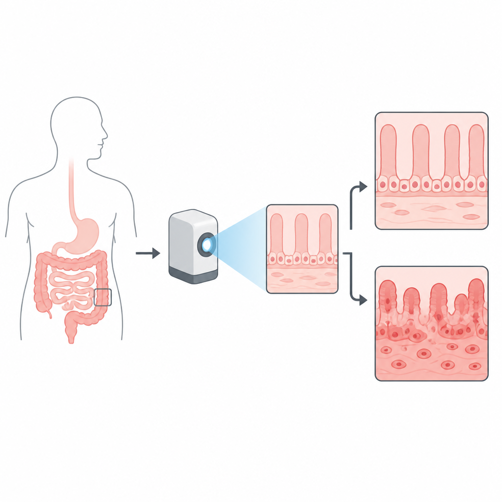
The study centers on optical coherence tomography, a technique that uses reflections of near infrared light to build a tiny three dimensional map of tissue structure, somewhat like ultrasound but with light instead of sound. Modern systems can resolve features on the scale of individual cells within about one to two millimeters of depth, enough to capture the important layers of the intestinal wall. The authors asked whether a high resolution version of this method could clearly show the normal patterns of the small intestine and colon and how those patterns change in inflammatory bowel disease.
From Operating Room to Imaging Bench
Tissue samples came from patients undergoing bowel surgery who had either healthy looking segments or areas affected by Crohn’s disease or ulcerative colitis. After removal, small pieces of bowel were cooled in solution, mounted flat beneath a glass coverslip, and immersed in a clear salt solution for imaging. A custom system using light centered at 1300 nanometers scanned each sample in a grid to build three dimensional data blocks with single micron scale detail. The same pieces were then processed in the usual way for lab slides, stained, and digitized so that the light based images could be directly compared with standard pathology.
Healthy Patterns Versus Diseased Patterns
In healthy small intestine, the team saw a forest of finger like projections when looking from above and from the side, matching the villi that increase surface area for absorption. In healthy colon, they saw orderly, tube like crypts arranged in a tight, regular grid with similar size and spacing, as well as a thin bright band marking the muscle layer beneath the lining and a looser supporting layer below that. These same features appeared in the usual stained slides, confirming that the light based method was truly capturing known anatomy in three dimensions.
How Crohn’s and Colitis Disturb the Landscape
In samples from people with Crohn’s disease, the usual order was disrupted. In the small intestine, villi appeared less distinct and more jumbled. In the colon, crypts varied in width and height and were less tightly packed, and thick horizontal bands of brighter tissue appeared beneath the lining, likely reflecting scarring in the supporting layer. In ulcerative colitis, the colon lining looked even more chaotic, with distorted crypts, pockets of trapped material called abscesses, and a thickened surface layer that blocked the view of deeper layers. These changes matched what pathologists saw in the stained sections, showing that the three dimensional light images tracked the same disease features.
Turning Texture into Numbers
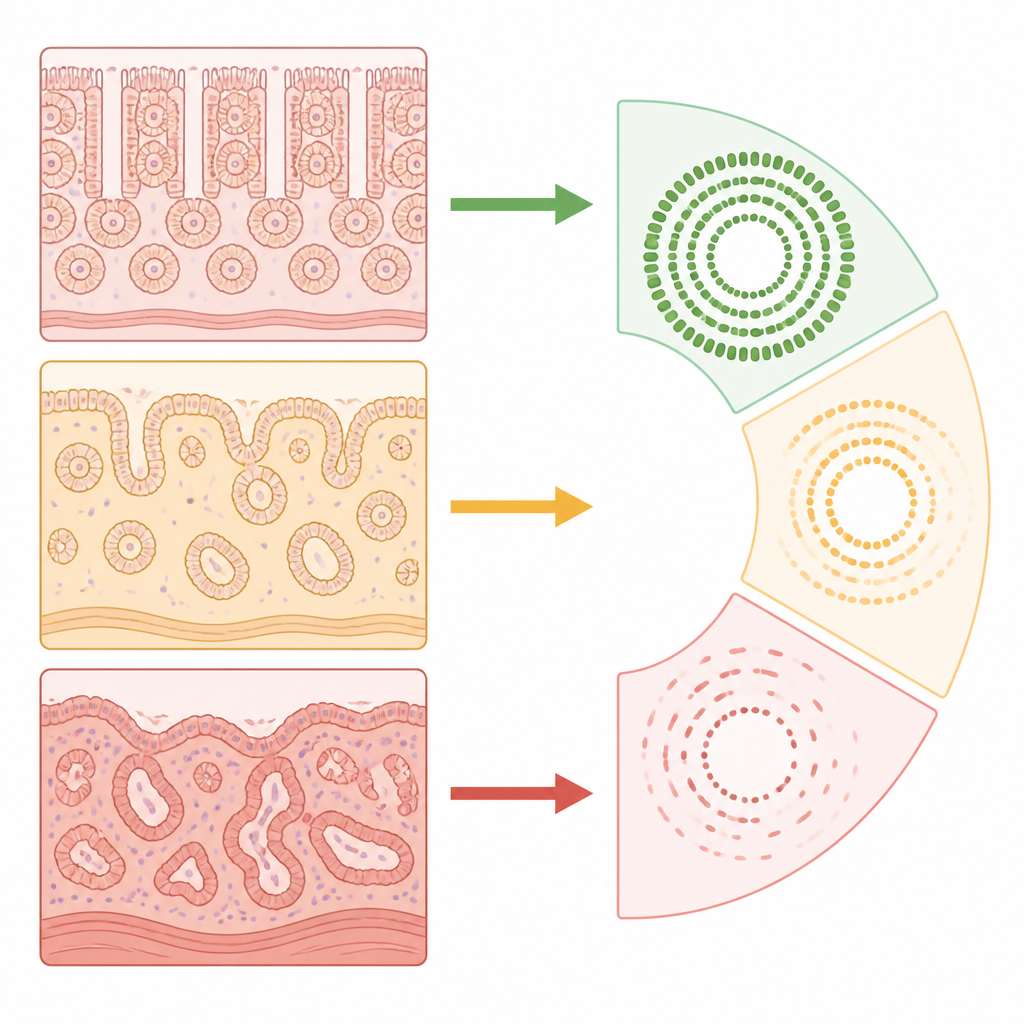
Beyond visual impressions, the researchers used a mathematical tool to describe how regular or irregular the surface patterns were in the colon. They analyzed top down views of the lining and converted the repeating crypt pattern into a map of spatial frequencies. Normal tissue produced a clear ring shaped signal, reflecting its even spacing, while inflamed samples from both Crohn’s disease and ulcerative colitis lost this ring because their crypts were distorted and uneven. By measuring the strength of this ring, the team could reliably distinguish normal from inflamed tissue, suggesting a possible numerical marker for disease involvement.
What This Could Mean for Patients
Overall, the study shows that high resolution optical coherence tomography can capture gut tissue architecture in three dimensions, revealing both fine details and broader patterns that differ between normal and inflamed bowel. While the work was done on tissue removed during surgery and the method still faces limits in how deep it can see, it lays groundwork for future tools that might scan the bowel wall during routine procedures, helping doctors gauge inflammation and scarring without extra biopsies. For patients, that could one day mean clearer answers about disease type and extent using gentler, more informative imaging.
Citation: Matt, A., Li, Y., Song, A. et al. Three-dimensional ex-vivo visualization of normal and inflamed small intestine and colonic tissue using optical coherence tomography. Sci Rep 16, 15568 (2026). https://doi.org/10.1038/s41598-026-46293-4
Keywords: inflammatory bowel disease, Crohn’s disease, ulcerative colitis, optical coherence tomography, intestinal imaging