Clear Sky Science · en
AAV.PHP.eB-based strategies for precise modulation of α7 nicotinic acetylcholine receptor in neurons and astrocytes in the adult mouse brain
Why tuning brain switches matters
Inside the brain, tiny protein “switches” help control how nerve cells talk to each other and how the brain responds to injury and disease. One of these switches, called the alpha7 nicotinic receptor, is linked to memory, attention, and inflammation. When it is too active or too quiet, it has been associated with conditions such as schizophrenia, Alzheimer’s disease, and problems after stroke. This study describes a toolkit to turn that specific switch up or down only in selected brain cells in adult mice, opening the door to more precise experiments and, eventually, more targeted therapies.
A shared switch in two kinds of brain cells
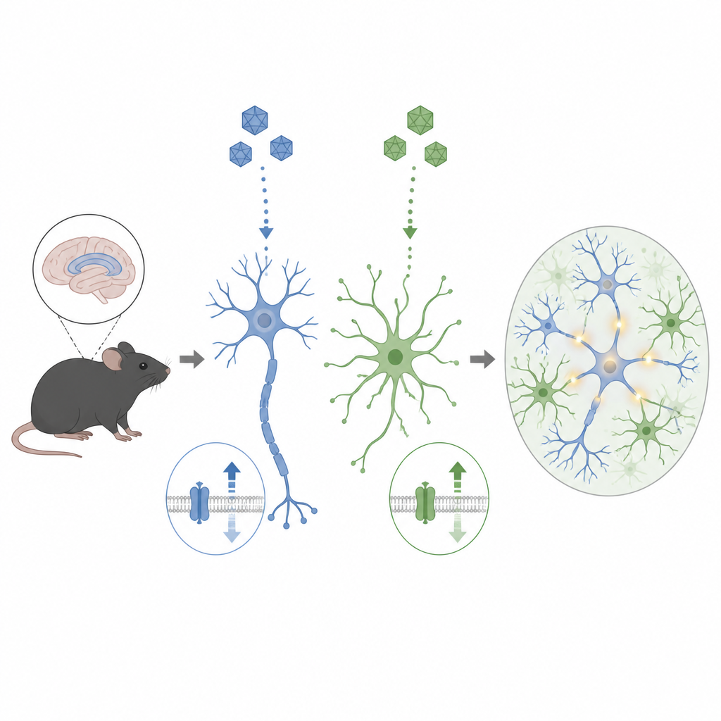
The alpha7 receptor is a protein channel that lets calcium ions flow into cells when activated. It is found not only on neurons, which send electrical signals, but also on astrocytes, support cells that shape brain chemistry and inflammation. Because these two cell types play different roles, scientists need tools that can change the receptor level in one cell type without disturbing the other. Until now, such fine control in the living brain has been hard to achieve, limiting efforts to understand how the receptor contributes to learning, memory, and immune responses in health and disease.
Building a targeted genetic toolkit
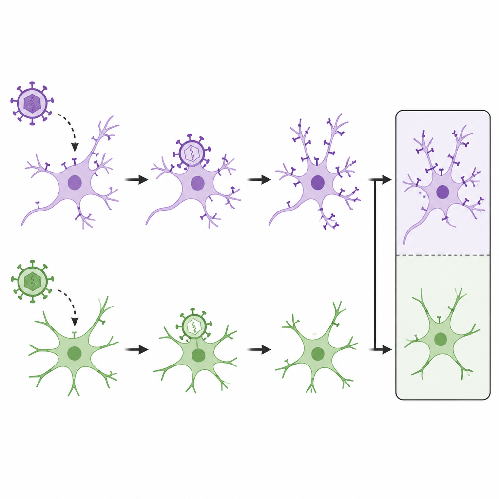
The researchers engineered a set of harmless viral carriers, based on an adeno associated virus variant called AAV.PHP.eB, to either boost or reduce the alpha7 receptor in specific cells. They inserted DNA “promoters” that act like address labels: one (called hSyn) directs the virus to work mainly in neurons, and another (called GFAP) directs it to astrocytes. For turning the receptor up, they packed in an extra copy of the receptor gene. For turning it down, they designed short RNA hairpins that trigger the cell to break down its own receptor message. Each construct also carried a fluorescent marker, so infected cells could be seen under the microscope.
Testing the tools from dish to living brain
The team first checked their designs in cultured human like cells and mixed mouse brain cells grown in dishes. They showed that the “overexpression” constructs raised receptor gene levels by several orders of magnitude, while the best RNA hairpin sequences sharply reduced those levels and weakened calcium signals triggered through the receptor. They then moved to more realistic models: brain slice cultures and, finally, adult mice. After injecting the viral vectors into the hippocampus, a region important for memory, they found that neuron targeted viruses lit up and altered receptor levels mainly in neurons, while astrocyte targeted versions did so mainly in astrocytes. Protein measurements confirmed strong, selective changes in the receptor, and there was little evidence of infection in off target cells.
Checking safety and cell reactions
One concern with viral delivery to the brain is that it might irritate or damage tissue, especially by activating astrocytes, which can swell and form scars. To address this, the scientists measured levels of GFAP, a protein that rises when astrocytes become reactive. Across cell cultures and brain samples from injected mice, they saw no significant increase in this marker compared with controls. This suggests that, under the tested conditions, their AAV.PHP.eB based vectors were well tolerated and did not trigger noticeable inflammation or scarring in the hippocampus.
What this means for future brain research
In plain terms, this work delivers a set of precise knobs for turning a key brain receptor up or down, separately in neurons and astrocytes, in the adult mouse brain. Researchers can now use these tools to tease apart how the alpha7 receptor shapes communication between cells, influences memory and attention, and modulates brain inflammation. In the longer term, the same strategy could help test whether restoring a healthy level of this receptor might ease symptoms in disorders where it is disrupted. While this study does not test treatments directly, it lays the technical groundwork for more targeted, cell specific approaches to brain disease.
Citation: Puliatti, G., Renna, P., Battistoni, M. et al. AAV.PHP.eB-based strategies for precise modulation of α7 nicotinic acetylcholine receptor in neurons and astrocytes in the adult mouse brain. Sci Rep 16, 15439 (2026). https://doi.org/10.1038/s41598-026-46279-2
Keywords: alpha7 nicotinic receptor, astrocytes, neurons, AAV gene delivery, mouse hippocampus