Clear Sky Science · en
Labeling and imaging of neurons with a genetically encoded autonomous bioluminescence system
Why glowing brain cells matter
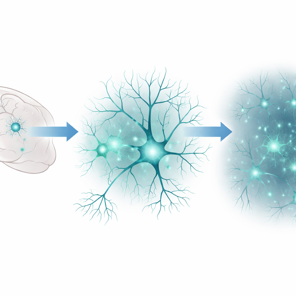
Watching living brain cells over days or weeks is essential for understanding how learning happens and how diseases like Alzheimer’s or Huntington’s slowly damage the nervous system. Yet most common imaging methods rely on bright external light that can itself harm delicate neurons and cause the fluorescent tags inside them to fade. This study presents a way for neurons to make their own gentle light continuously, without any added chemicals, allowing scientists to track their health and behavior in real time.
A new way for cells to light themselves up
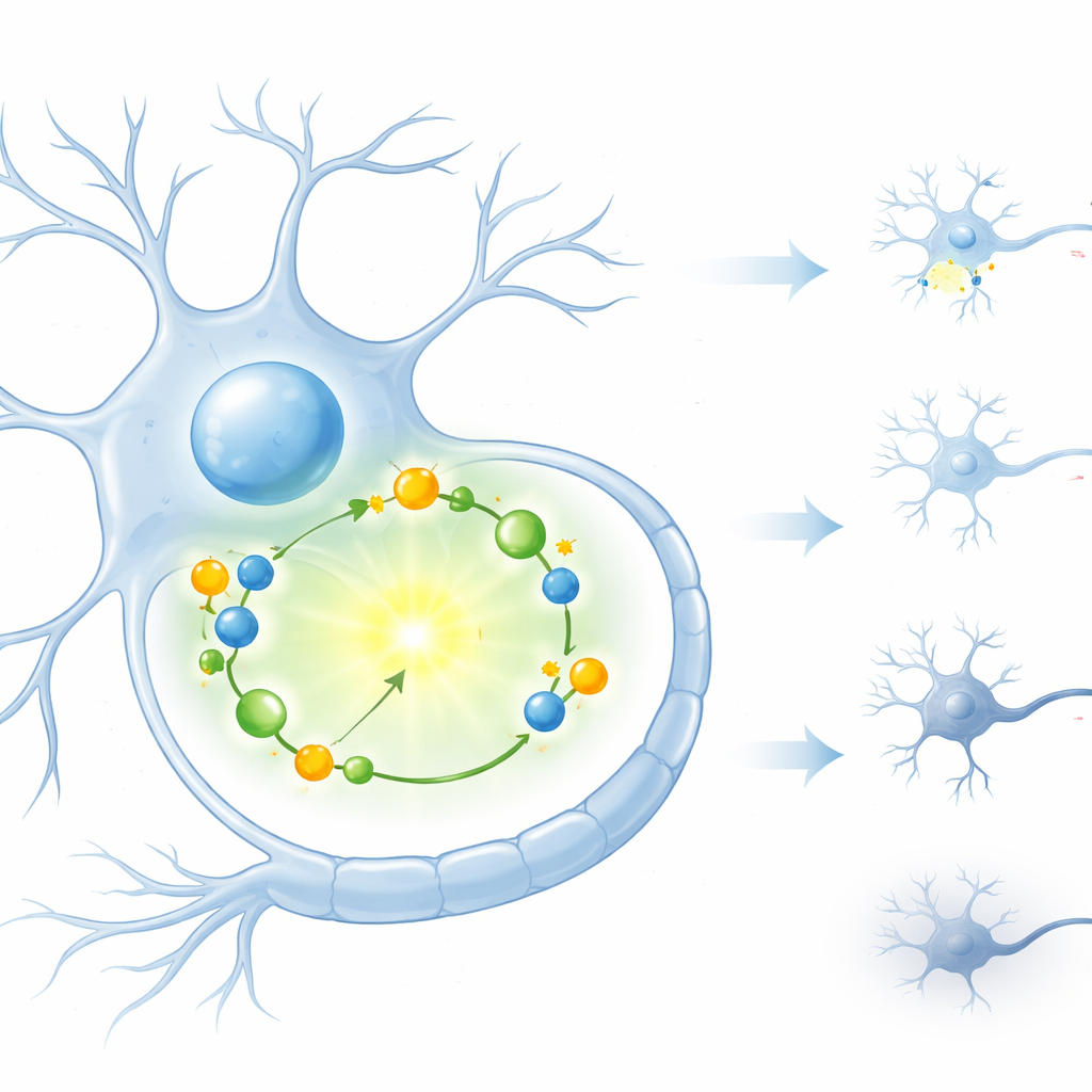
The researchers tapped into a natural light-making system originally found in bacteria. In those microbes, a group of proteins work together to produce a steady glow through a chemical reaction. Instead of shining light from the outside, the cell itself drives the reaction from the inside, so no external illumination is needed. Importantly, this light is only produced when the cell’s internal energy supply is intact, tying the glow directly to cellular health. Bringing this multi-part system into fragile mammalian neurons, however, is technically challenging because it requires six different genes that all have to be delivered and switched on together.
Smuggling a six-part light kit into neurons
To solve this problem, the team used adeno-associated viruses—small, well-studied delivery vehicles often used in gene therapy—to carry the bacterial light-making genes into mouse neurons. They first tested different genetic “on switches” to find one that would drive strong expression in nerve cells. Then they combined this powerful switch with a neuron-specific control system so that only neurons, and not surrounding cell types, would light up. Because each virus can hold only a limited amount of genetic material, the six bacterial genes had to be smartly split and paired across several viral vectors. By experimenting with different groupings, the researchers identified a combination of four virus types that produced a bright, stable glow while still fitting within the packaging limits.
Seeing single neurons and sensing their health
Once optimized, the viral mix—referred to as Lux AAVs—allowed neurons to continuously glow strongly enough to be imaged one cell at a time with sensitive cameras. The brightness reached roughly one-third of that of a widely used firefly-based system, but with a key advantage: the cells did not need repeated doses of an external light-producing chemical. The glow from Lux-labeled neurons remained stable for at least 20 hours, enabling long-term, hands-off observation. Crucially, tests showed that running this light system did not noticeably harm the cells, despite the fact that it consumes some of their energy resources.
Watching cells decline under stress and disease-like conditions
Because the bacterial light reaction depends on the cell’s energy molecules, the team asked whether fading light could act as an early warning signal of trouble. They exposed Lux-labeled neurons to hydrogen peroxide to induce oxidative stress, a damaging condition linked to many neurodegenerative diseases. Over several hours, the glow gradually diminished and then disappeared, matching what is known about loss of neuron viability under such stress. Next, they bathed neurons in high levels of glutamate, a brain signal molecule that in excess becomes toxic. Here, the light dropped quickly to a fraction of its starting level and then partially rebounded over the next day, suggesting a mix of reversible energy disruption and lasting cell damage. Finally, they mimicked Huntington’s disease by forcing neurons to produce a mutant form of the huntingtin protein that forms toxic clumps. Cells expressing this harmful version showed about a quarter less light than control cells, revealing measurable stress even before outright cell death.
What this means for future brain research
This work delivers a practical toolkit for making neurons autonomously glow in a way that directly reflects their metabolic health. Because the light is generated from within and does not require external flashes or repeated chemical additions, researchers can watch the same cells continuously over long periods. That makes it easier to detect the earliest signs of stress, follow how damage unfolds, and test whether potential treatments can preserve or restore cellular vitality. In simple terms, the authors have turned neurons into tiny self-reporting lanterns, offering a powerful new window into how brain cells live, struggle, and die in the face of disease.
Citation: Brinker, T., Günther, A., Kiszka, K.A. et al. Labeling and imaging of neurons with a genetically encoded autonomous bioluminescence system. Sci Rep 16, 12892 (2026). https://doi.org/10.1038/s41598-026-46211-8
Keywords: neuronal imaging, bioluminescence, gene delivery, neurodegeneration, cell health monitoring