Clear Sky Science · en
Comparative analysis of different biophysical techniques for exosome characterization
Tiny Messengers With Big Potential
Inside our bodies, cells are constantly talking to each other using tiny bubbles called exosomes. These nanoscale packets can carry proteins and genetic material, and researchers hope to harness them as natural delivery vehicles for future medicines, especially in cancer treatment. But before exosomes can be safely used as drug carriers, scientists need reliable ways to measure their size, purity, and concentration—no easy task for particles far smaller than the wavelength of light.
Why Measuring These Bubbles Is So Hard
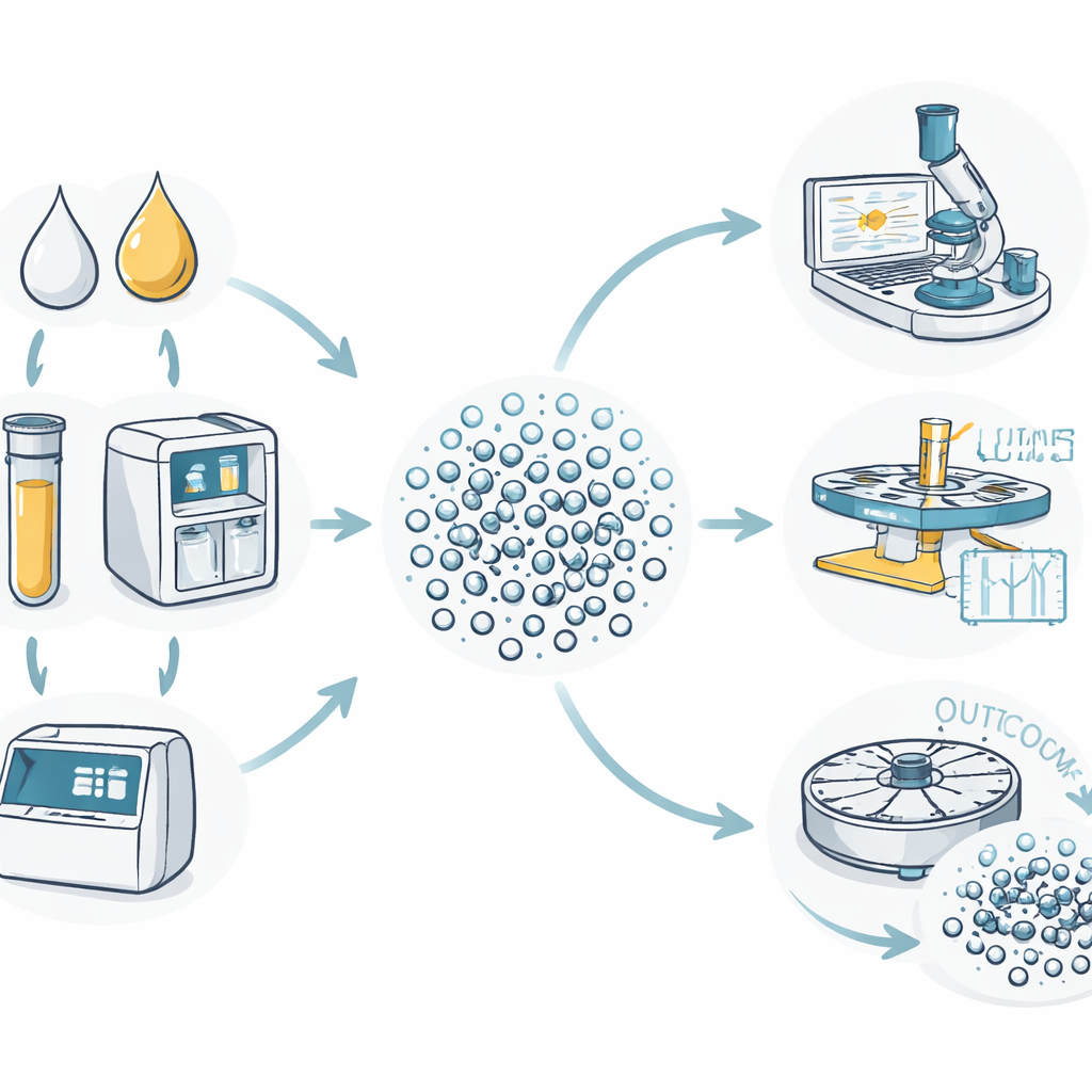
Exosomes are incredibly small—about one‑thousandth the width of a human hair—and they rarely travel alone. In milk or urine, they mingle with proteins, fats, and other microscopic debris. Different measuring tools can easily give different answers for how big they are, how many are present, and how clean a sample really is. The team behind this study set out to compare several widely used physical measurement methods on the same sets of exosomes, purified from cow’s milk and human urine by either traditional high‑speed spinning (ultracentrifugation) or a newer automated system called EXODUS. Their goal was not just to count and size these particles, but to see which techniques are best suited for which questions.
Fast First Glance Versus Detailed Tracking
One common tool, dynamic light scattering, shines a laser through a sample and analyzes how the scattered light flickers as particles jiggle in water. It is quick and gentle, making it useful for screening large numbers of samples. However, the authors found that even a few larger contaminants can strongly distort the signal, because big particles scatter light much more intensely than small ones. In their milk and urine samples, this method often reported broad size ranges and inconsistent results when the samples were mixed in size, showing that it works best only when the particles are already fairly uniform and clean.
Watching Single Particles One by One
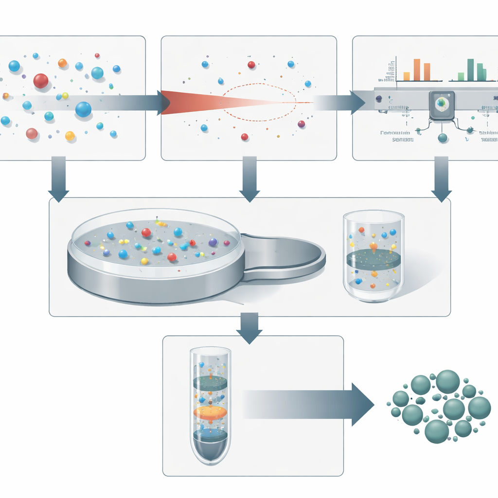
Nanoparticle tracking analysis takes a different approach: it watches individual particles move under the microscope and calculates their sizes from their motion. This single‑particle view gave a sharper picture of how exosome sizes differed between purification methods. Samples processed by EXODUS tended to contain smaller and more evenly sized particles than those obtained by simple ultracentrifugation, suggesting fewer impurities. The technique could also clearly reveal changes caused by filtering or freezing and thawing, although leftover contaminants still nudged the measured sizes above the textbook range for exosomes.
Counting, Purity Checks, and Hidden Impurities
A third method, NanoCoulter, measures tiny changes in electrical resistance as each particle squeezes through a nanoscale pore. This allowed the researchers to obtain absolute particle counts and size distributions without relying on light. Within its working size window, it agreed well with electron microscope images and was less affected by small debris than optical methods. However, its chip could not detect the very smallest impurities, and it was less sensitive to subtle shifts in size after processing steps. The final technique, analytical ultracentrifugation, used very high‑speed spinning combined with ultraviolet light detection to follow how different components settle. By comparing signals that reflect proteins versus genetic material, the authors could see clear signatures of protein contaminants in milk exosome preparations and how additional cleaning steps or EXODUS removed them.
Why No Single Tool Is Enough
Together, these tests painted a consistent picture: each method reveals part of the story but has blind spots. Light scattering is fast but easily misled by a few large particles. Particle tracking offers fine detail but can be skewed by leftover debris. Electrical sensing excels at counting and sizing within a specific range yet misses very small impurities. High‑speed spinning with optical readout is powerful for judging purity and separating exosomes from proteins, but it is complex and less straightforward for precise sizing. The authors conclude that building a trustworthy, standardized way to characterize exosomes—essential for turning them into dependable drug carriers—will require combining multiple complementary techniques rather than relying on any single favorite instrument.
Citation: Yu, X., Wang, Z., Zhang, R. et al. Comparative analysis of different biophysical techniques for exosome characterization. Sci Rep 16, 10724 (2026). https://doi.org/10.1038/s41598-026-46079-8
Keywords: exosomes, drug delivery, nanoparticle analysis, biophysical techniques, sample purity