Clear Sky Science · en
Discordance between vibration controlled-transient elastography and ultrasound in cirrhosis assessment: prognostic implications for liver-related events in patients with chronic hepatitis B
Why spotting hidden liver damage matters
People living with chronic hepatitis B often feel well for years, yet quiet damage can build up in the liver and lead to scarring, cirrhosis, and liver cancer. Doctors rely on noninvasive scans to catch this damage early, but different tests do not always agree. This study asks what happens to patients when a standard ultrasound scan and a newer stiffness test give conflicting answers about whether cirrhosis is present, and which result better predicts future liver problems.
Two common tools to look inside the liver
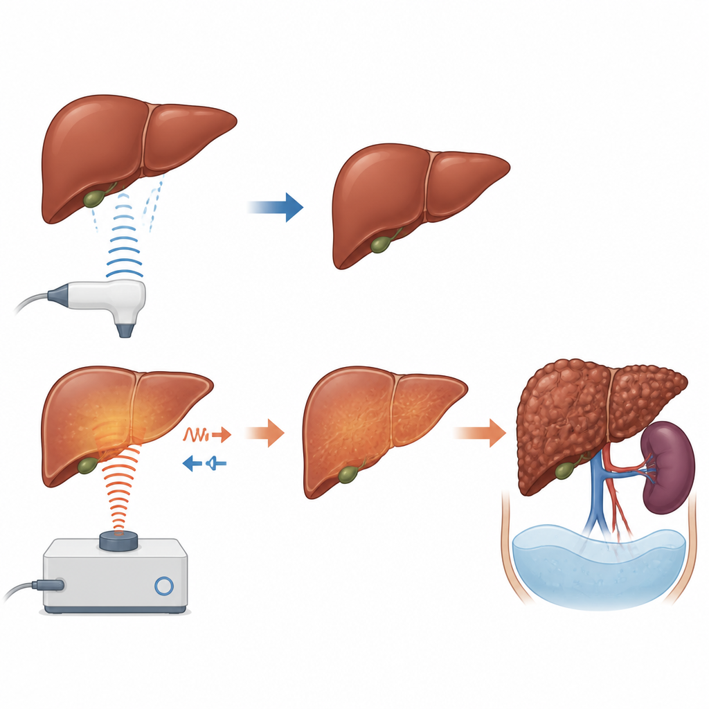
Today, most patients with chronic liver disease are monitored using two painless tests. Traditional ultrasound creates real-time images of the liver and nearby organs, allowing radiologists to look for a bumpy surface, altered shape, or enlarged spleen that hint at cirrhosis. Vibration-controlled transient elastography, often known by the device name FibroScan, works differently. It sends a gentle vibration through the liver and measures how fast the wave travels, which reflects how stiff the liver tissue is. Stiffer livers are usually more scarred. Because liver biopsy is invasive and risky, these scans are widely used to guide treatment and cancer surveillance.

When test results do not match
The researchers reviewed records from 532 adults with chronic hepatitis B treated at a university hospital in southern Thailand between 2014 and 2021. None had already developed liver cancer or serious cirrhosis complications at the start. Each had both ultrasound and elastography around the same time. The team defined cirrhosis on elastography as a liver stiffness value of at least 10 kilopascals, following international guidance, and used radiologists’ written reports to classify cirrhosis on ultrasound. This allowed them to place patients into four groups: both tests saying no cirrhosis, only ultrasound saying cirrhosis, only elastography saying cirrhosis, or both tests agreeing on cirrhosis.
Tracking who developed serious liver problems
The patients were followed for a median of 4.4 years to see who went on to develop liver cancer or major complications of cirrhosis, such as fluid in the abdomen, internal bleeding, or confusion from liver failure. Most patients (about four out of five) had no cirrhosis according to either test and very few of them developed problems. A smaller group had clear cirrhosis on both tests, and these patients had by far the highest rate of serious events over five years. Strikingly, people whose ultrasound looked non-cirrhotic but whose elastography showed a stiff liver had a clearly higher risk than the no-cirrhosis group, even though their scans looked reassuring by standard imaging. In contrast, those labeled cirrhotic by ultrasound alone, but with low stiffness values, did not have more events than the clearly non-cirrhotic patients.
Checking the findings from different angles
To make sure the pattern was not a fluke, the team repeated their analyses using stricter definitions. They raised the stiffness threshold to 12.5 kilopascals, as recommended by the World Health Organization, and also limited the sample to people with only mildly raised liver enzymes, which reduces the chance that temporary inflammation was falsely making the liver look stiff. In each case, patients with high stiffness but reassuring ultrasound findings still faced a higher risk of liver cancer or cirrhosis complications. These sensitivity checks suggest that elastography is capturing genuine underlying scarring rather than short-lived changes.
What this means for patients and doctors
For a person with chronic hepatitis B, these results highlight that a normal-looking ultrasound does not always mean the liver is safe. A stiff liver on elastography, even when the ultrasound image appears mild, signals a higher chance of serious liver trouble in the coming years and may justify closer follow-up and carefully planned treatment. At the same time, an ultrasound that suggests cirrhosis without a matching rise in stiffness may sometimes overcall disease. The study does not replace the need for expert judgment or, in uncertain cases, a biopsy. But it shows that using liver stiffness measurements alongside ultrasound can sharpen risk assessment, helping clinicians focus attention on those most likely to benefit from intensive monitoring while sparing others from unnecessary worry and testing.
Citation: Uman, N., Chamroonkul, N., Kaewdech, A. et al. Discordance between vibration controlled-transient elastography and ultrasound in cirrhosis assessment: prognostic implications for liver-related events in patients with chronic hepatitis B. Sci Rep 16, 15645 (2026). https://doi.org/10.1038/s41598-026-45880-9
Keywords: chronic hepatitis B, liver cirrhosis, ultrasound, transient elastography, hepatocellular carcinoma