Clear Sky Science · en
Enhancing lung diseases recognition through CNN-RNN methodologies
Why smarter lung scans matter
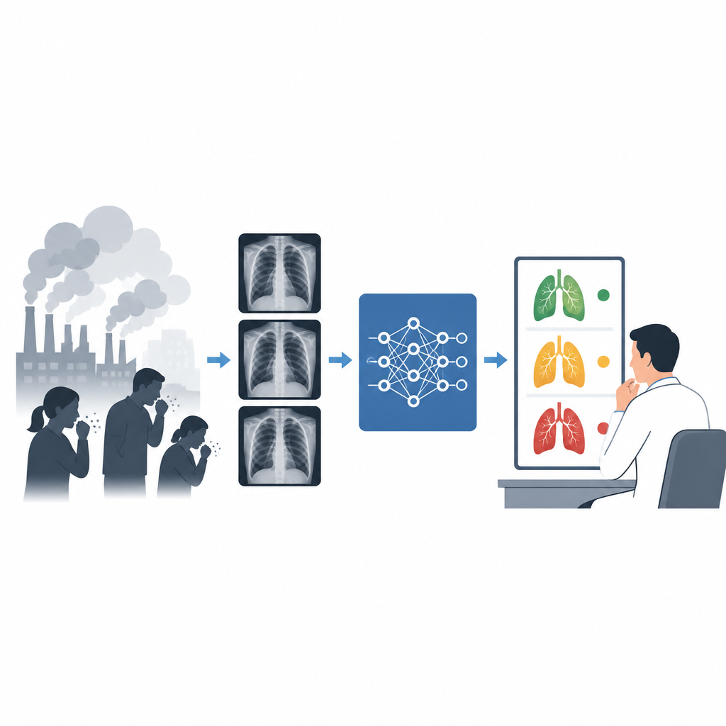
Respiratory illnesses like pneumonia, COVID, and tuberculosis kill millions of people each year, especially in low income countries where doctors and scanning equipment are in short supply. Chest X rays are cheap and widely available, but they can be hard to interpret and different lung diseases often look similar. This study explores how a compact form of artificial intelligence can help spot several kinds of lung problems in X ray images more quickly and reliably, without replacing the judgement of trained clinicians.
The global burden on lungs
The authors begin by describing how lung disease has become a leading cause of death worldwide, with a particularly heavy toll in Bangladesh. Pollution, dust, smoke, and infections contribute to rising rates of pneumonia, asthma, tuberculosis, influenza, COVID, and other chronic breathing disorders. Symptoms such as cough, chest pain, and shortness of breath overlap, making it difficult to tell one condition from another based on clinical signs alone. At the same time, many regions lack enough radiologists to read X rays, and delays in diagnosis can be deadly, especially for young children and older adults with other health problems.
Turning X rays into patterns a computer can read
Because X ray machines are far more common and affordable than CT scanners, the study focuses on automating the reading of chest X rays. The researchers use a large public collection of more than twenty thousand images labelled as COVID 19, lung opacity, normal lungs, or viral pneumonia. Before training their system, they carefully resize and normalize each image and then cut it into many small square patches. This step adds variety to the data and helps the model pay attention to fine details that might signal infection or fluid. The images are then split into training and testing sets so the system can be evaluated on X rays it has never seen before.
Two digital eyes on each image
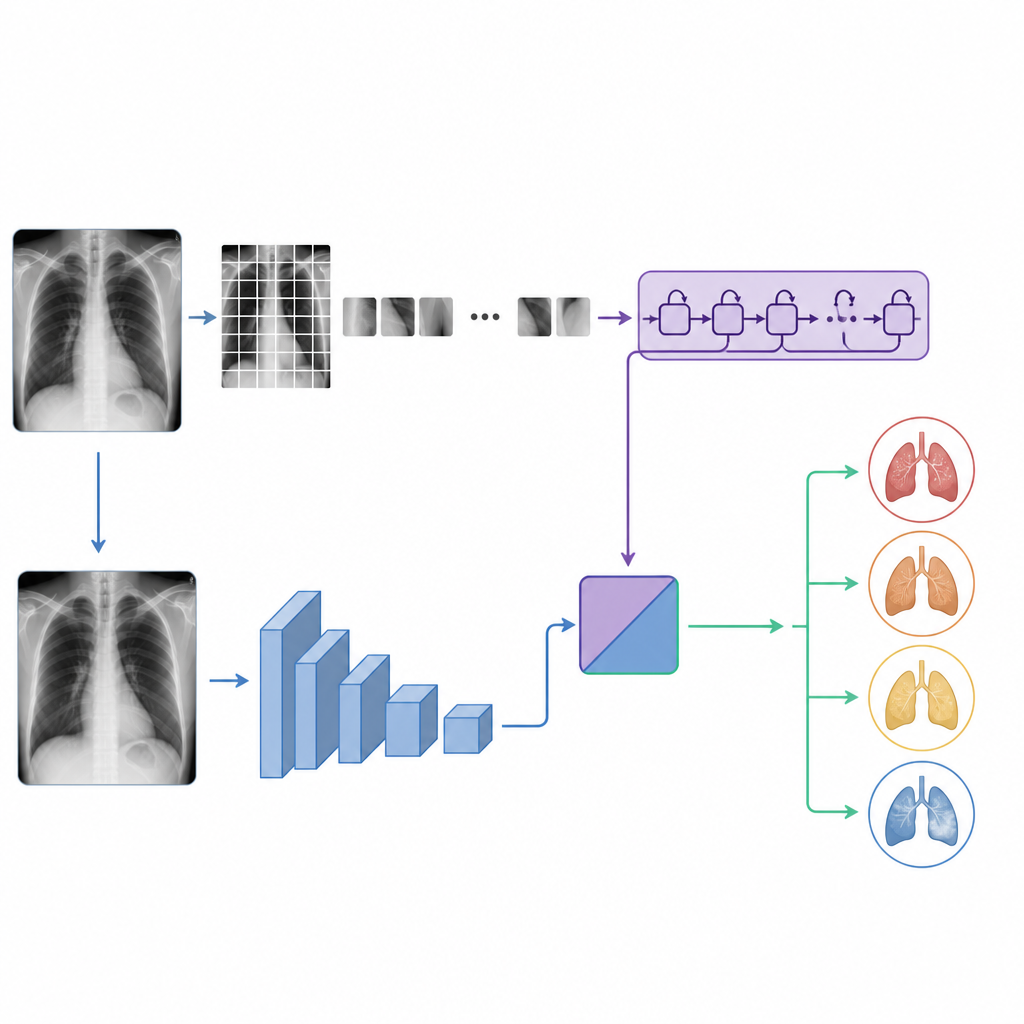
The core of the work is a new hybrid model called C RNet that combines two different kinds of neural networks. One branch is a conventional image recognizer that scans the whole X ray to learn spatial patterns such as cloudy areas, sharp edges, and overall lung shape. The second branch treats the grid of patches as a sequence and uses a recurrent network to capture how small regions relate to one another across the image. By joining the outputs of these two branches, the system learns both local details and broader context, like how shadows in one part of the lung line up with changes elsewhere. A final layer turns this combined information into one of four possible diagnoses.
Seeing what the machine sees
To make the system more trustworthy for clinicians, the authors add an explainable AI tool called Grad CAM. This method overlays a colored heat map on top of the original X ray, highlighting the regions that most influenced the model’s decision. In tests, C RNet tends to focus on realistic disease areas within the lung fields, while comparison models sometimes light up irrelevant regions near the shoulders or diaphragm. This visual feedback can help radiologists judge whether the algorithm is keying in on meaningful anatomy or being distracted by noise or artifacts in the image.
How well the system performs
When evaluated on the full dataset, C RNet correctly classified lung condition in about 94 out of 100 cases, outperforming several well known deep learning models such as Xception, VGG19, a vision transformer, and a CNN linked to a recurrent layer in a simpler way. It was particularly strong at recognizing COVID and viral pneumonia while still distinguishing normal lungs and more subtle lung opacity cases. Importantly, the model achieves this accuracy with fewer than two million trainable parameters and a file size of about seven megabytes, making it far smaller than many popular image recognition networks. This compact design means it can run on modest computers or hospital machines without specialized hardware.
What this means for patients and clinics
For a lay audience, the main message is that the study shows how a smart but lightweight computer program can help doctors read chest X rays more consistently. By combining two ways of looking at the same image and then showing which parts of the lungs drove its decision, C RNet offers both accuracy and transparency. In resource limited settings, such a tool could serve as an extra pair of eyes, flagging suspicious cases for closer review and helping ensure that serious lung infections are detected earlier. While it does not replace medical expertise, it has the potential to support faster, more reliable diagnoses where they are needed most.
Citation: Zahin, I.A., Ahsan, M.F., Orni, R.A. et al. Enhancing lung diseases recognition through CNN-RNN methodologies. Sci Rep 16, 15819 (2026). https://doi.org/10.1038/s41598-026-45842-1
Keywords: lung disease, chest X ray, deep learning, medical imaging AI, pneumonia detection