Clear Sky Science · en
Multi-class classification of brain tumor using a ResNet101 backbone integrated with multi-scale deformable attention module and advanced data augmentations
Why brain scan sorting matters
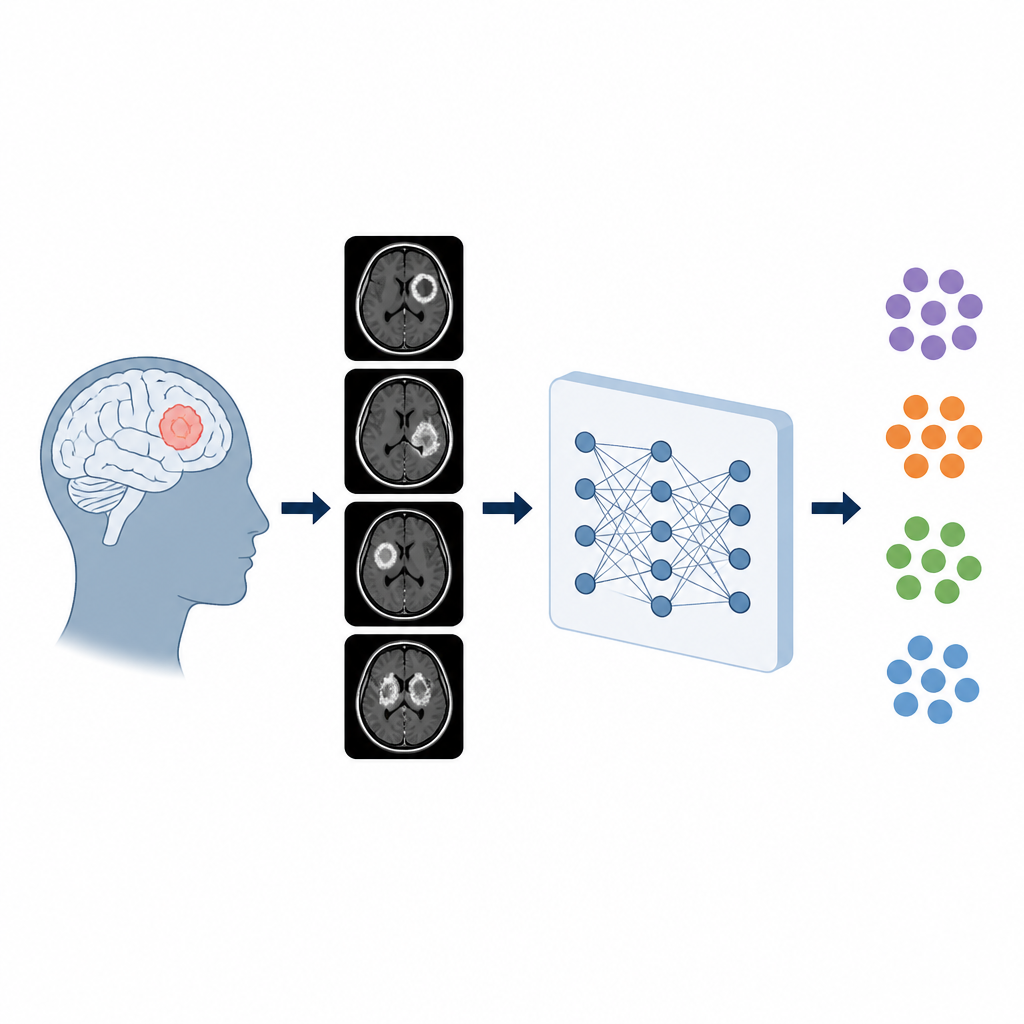
When doctors look at brain scans to check for tumors, they must determine not only whether a tumor is present but what type it is. Different tumor types call for different treatments, yet they can look strikingly similar on Magnetic Resonance Imaging (MRI). This study explores a new artificial intelligence approach that can sort many kinds of brain tumors from MRI scans with very high accuracy, potentially giving radiologists faster and more consistent support when they make life-impacting decisions.
Teaching computers to read brain scans
The researchers focus on multi-class classification, which means telling apart many tumor categories at once rather than just spotting tumor versus no tumor. They use a deep learning model built on ResNet101, a widely used image-recognition network that has already learned general visual patterns from large natural-image collections. The team adapts this network to brain MRI and applies it to a public dataset that includes 15 classes, from normal brain tissue to specific tumors such as glioblastoma, meningioma, and medulloblastoma. Tackling so many categories makes the task much closer to what radiologists face in real clinics.
Cleaning and balancing real-world data
Medical image collections often contain hidden pitfalls, such as duplicate scans of the same patient or large differences in how common each tumor type is. To avoid misleading results, the authors first scan the entire dataset, compute a digital fingerprint for every image, and remove exact duplicates. They then group images by patient ID and split patients, not images, into training, validation, and test sets so that the same person’s scans never appear in more than one subset. This patient-wise split helps ensure that reported accuracy reflects performance on truly unseen individuals rather than repeated views of the same brain. The team also uses a battery of image transformations and a technique called MixUp to make rare tumor types better represented during training and to help the model cope with variations in orientation, brightness, and noise.
Looking closer with flexible attention
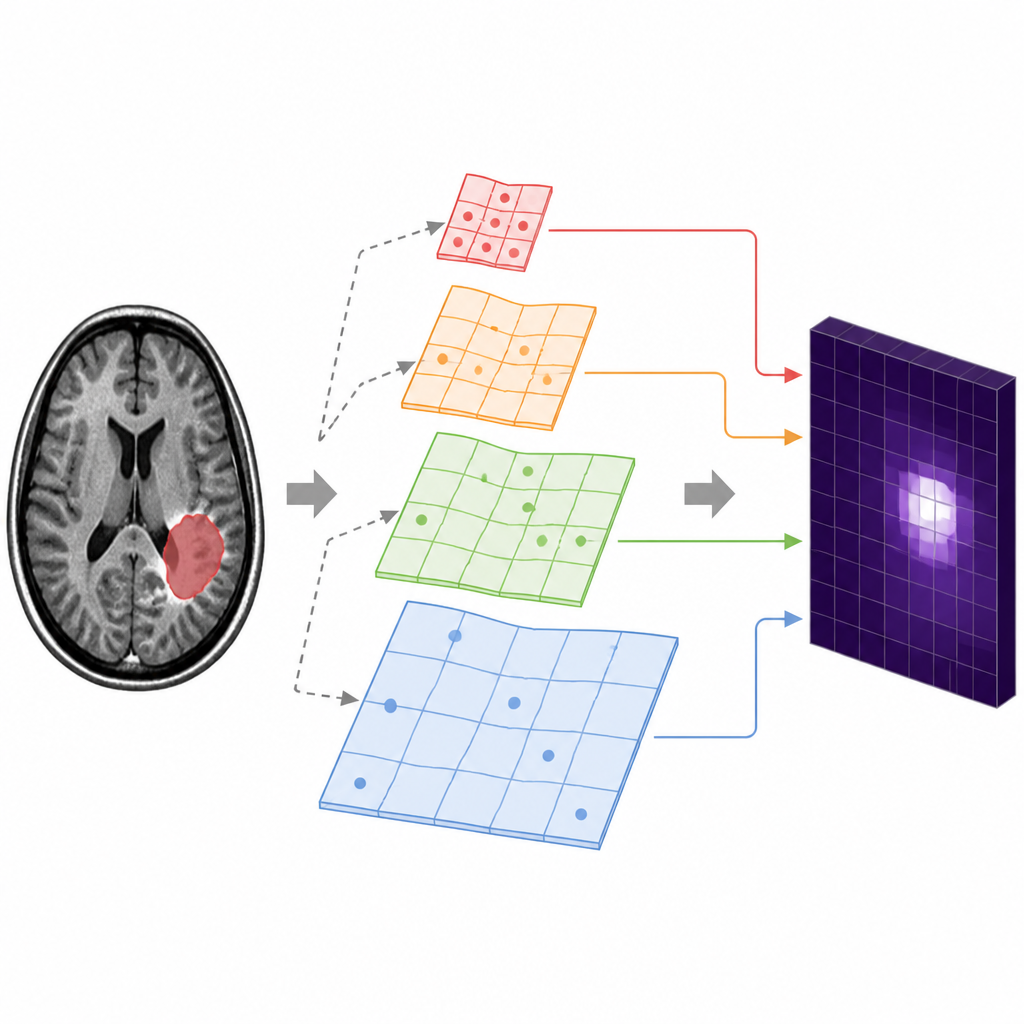
A key innovation in the study is a module the authors call a multi-scale deformable attention module. In simple terms, the model examines the MRI at different levels of detail, from coarse structures of the whole brain down to finer textures near the tumor. Instead of scanning every location in a rigid, grid-like way, the deformable attention learns to slightly shift where it “looks” so it can zero in on informative regions. It then applies two complementary forms of attention: one that highlights important spots in the image and another that strengthens the most useful feature channels inside the network. This combination allows the system to better capture the subtle shape and texture differences that distinguish one tumor type from another.
Putting performance and trust to the test
After training for many cycles, the model achieves a validation accuracy of about 97 percent and a test accuracy above 99 percent across all 15 classes. Most tumor types are classified perfectly in the held-out test set, with only a handful of mistakes in tumor categories that are both rare and visually similar. The researchers compare their results with a prior method that also uses deformable attention and show that their approach matches or surpasses it on nearly all evaluation measures, despite the complex class mix. They further stress-test the system by adding noise and reducing image resolution, finding that accuracy remains high even when scan quality degrades, which is important for real-world hospital conditions.
Seeing what the model sees
To help clinicians trust the system, the authors use visualization tools that reveal where the network is focusing when it makes a decision. Heatmaps produced by Grad-CAM highlight areas on the MRI that most influenced the prediction, and SHAP-based analyses indicate which learned features push the model toward one diagnosis or another. These visual explanations consistently point to tumor regions and their boundaries rather than irrelevant background, suggesting that the model’s reasoning aligns with radiological intuition. While the study emphasizes that further external testing is needed, it demonstrates that a carefully designed deep learning pipeline can not only classify many brain tumor types with high reliability but also offer interpretable clues about how those decisions are made.
What this could mean for patients
In everyday clinical practice, this kind of system would not replace radiologists but act as a second reader, quickly flagging likely tumor types and drawing attention to suspicious regions on the scan. By handling a wide variety of tumors and remaining stable under noisy or lower-quality images, the framework described in this paper shows how AI could support more consistent and timely diagnoses. With additional validation across hospitals and scanners, such tools could become part of routine workflows, helping specialists focus on treatment planning while computers handle much of the repetitive image sorting.
Citation: Reddy, B.S., Jha, R.R., Dasore, A. et al. Multi-class classification of brain tumor using a ResNet101 backbone integrated with multi-scale deformable attention module and advanced data augmentations. Sci Rep 16, 15938 (2026). https://doi.org/10.1038/s41598-026-45675-y
Keywords: brain tumor MRI, deep learning, medical imaging AI, tumor classification, attention networks