Clear Sky Science · en
Cerebellar oligodendrocytic α-synuclein pathology and dentate nucleus neuronal hypertrophy in Parkinson’s disease
Why this brain study matters
Parkinson’s disease is usually linked to damage in a region deep in the brain that helps control movement. This study looks elsewhere, at a part of the “little brain” at the back of the head called the cerebellum, to ask whether overlooked cell types there may also be involved. By examining donated brains from people with Parkinson’s, the researchers uncover unexpected changes in support cells and neurons that could help explain both movement problems and some of the wider symptoms of the disease. 
A new look at Parkinson’s beyond movement centers
For many years, scientists have focused on the loss of dopamine producing nerve cells in the substantia nigra and the presence of clumps of a protein called alpha synuclein inside neurons, known as Lewy bodies. Yet people with Parkinson’s often have symptoms that go far beyond tremor and stiffness, including changes in thinking, mood, and automatic body functions. At the same time, modern brain mapping has revealed that the cerebellum and a key structure within it, the dentate nucleus, are tightly wired into circuits for both movement and higher mental functions. This raised the possibility that damage in cerebellar networks might contribute to the wide range of problems seen in Parkinson’s disease.
What the researchers examined in donated brains
The team studied cerebellar tissue from six people who had been diagnosed with idiopathic Parkinson’s disease and compared it with tissue from five people of similar age without known brain disease. They focused on the dentate nucleus and the white matter that carries its incoming and outgoing nerve fibers. Using a battery of microscopic staining techniques, they looked for alpha synuclein deposits, other common age related brain changes, the condition of the insulating myelin around nerve fibers, and the size of neurons in the dentate nucleus. Careful three dimensional measurements allowed them to estimate the volume of each cell body, its nucleus, and its nucleolus, a structure linked with protein production.
Hidden protein build up in support cells
A striking finding was the presence of alpha synuclein clumps inside oligodendrocytes, the support cells that form and maintain myelin, in the white matter around the dentate nucleus of every Parkinson’s case but none of the controls. These inclusions filled most of the visible cell body and looked different from classic Lewy bodies, which have a dense core and a pale halo and are found in neurons such as those of the substantia nigra. Only rare alpha synuclein positive nerve fibers were seen near the dentate nucleus, and notably no typical Lewy bodies were detected in dentate neurons themselves. Staining for myelin suggested a subtle pallor, hinting at thinning or loss of the protective sheath around nerve fibers in this region, though the authors note that future work with precise measurements will be needed to confirm this. 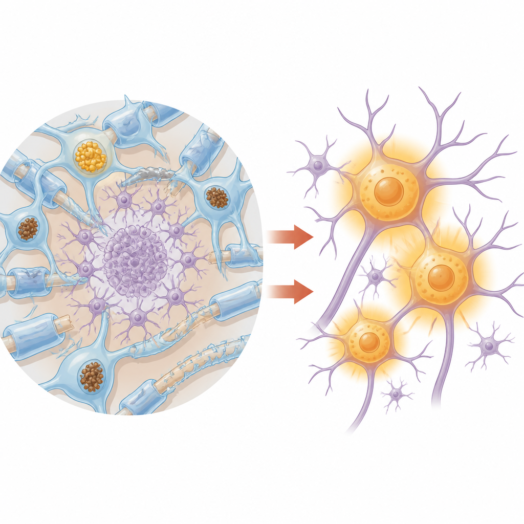
Overworked neurons in a stressed network
Although the neurons of the dentate nucleus did not contain Lewy bodies, they were not entirely normal. Stereology based measurements showed that, on average, their cell bodies, nuclei, and nucleoli were larger in the Parkinson’s group than in the control group. This enlargement, or hypertrophy, reached strong statistical significance, especially for the nuclear and nucleolar volumes. The authors interpret this pattern as a possible sign that these neurons are in a heightened metabolic state, working harder to compensate for stress or disrupted input and output along their connections. Because the dentate nucleus sends signals through the thalamus to many brain regions, including the basal ganglia, such stress could ripple through wider movement and cognitive networks.
What this could mean for people with Parkinson’s
Together, the findings suggest that in Parkinson’s disease, alpha synuclein does not only harm neurons in classic movement centers but also accumulates in myelin making support cells in the cerebellum. The authors propose that this “oligo synucleinopathy” in the dentate region may weaken the partnership between glial cells and neurons, disturb the flow of signals along cerebellar pathways, and contribute to both motor symptoms like tremor and balance problems and non motor issues such as cognitive or mood changes. While the study is small and cannot yet prove cause and effect, it widens the picture of Parkinson’s disease from a single damaged pathway to a more complex network disorder that involves both neurons and their supporting partners.
Citation: Iacono, D., Peng, H., Bouffard, J.P. et al. Cerebellar oligodendrocytic α-synuclein pathology and dentate nucleus neuronal hypertrophy in Parkinson’s disease. Sci Rep 16, 16199 (2026). https://doi.org/10.1038/s41598-026-45589-9
Keywords: Parkinson’s disease, cerebellum, alpha synuclein, oligodendrocytes, dentate nucleus