Clear Sky Science · en
FTIR imaging identifies alterations in lung tissue structure and biochemical composition in human idiopathic pulmonary fibrosis
Why lung scarring matters
Idiopathic pulmonary fibrosis is a serious lung disease in which the air sacs slowly turn into stiff scar tissue, making every breath harder. Doctors struggle to diagnose it early, and current treatments can only slow it down. This study explores a new, label free way to "see" chemical changes in lung tissue, in hopes of spotting the disease more clearly and perhaps earlier.
A chemical fingerprint of sick lungs
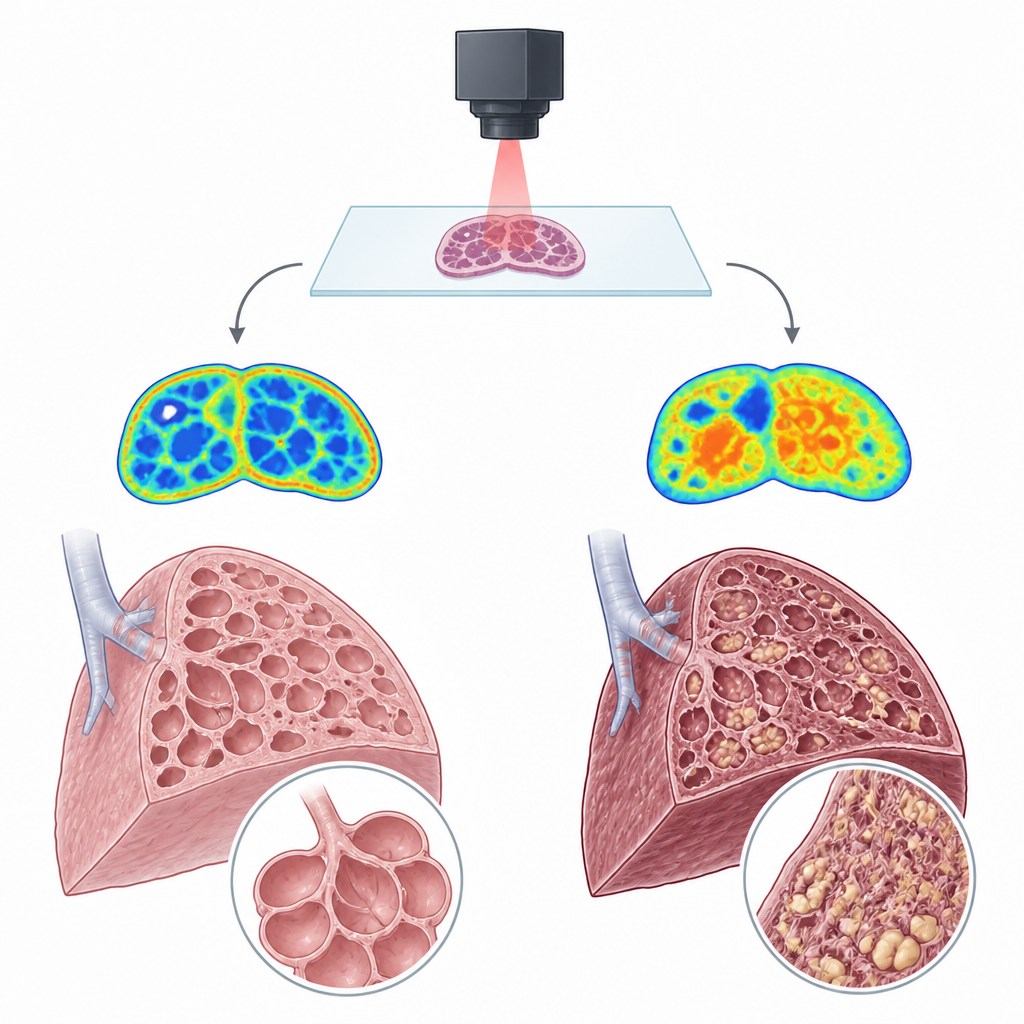
The researchers used a technique called Fourier transform infrared imaging, which shines invisible infrared light through thin slices of lung and records how different molecules absorb that light. Instead of adding dyes or labels, the method relies on the natural vibrations of fats and proteins in the tissue to create a chemical map. The team compared lung samples from people with idiopathic pulmonary fibrosis to samples from people without lung disease, carefully examining both heavily scarred and less affected regions.
Fats build up as lungs scar
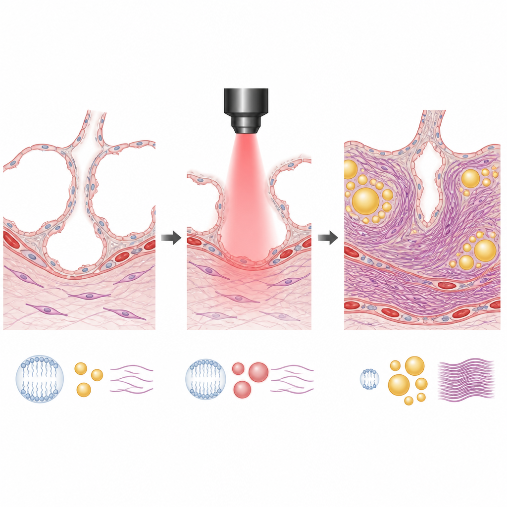
One of the clearest differences between diseased and healthy lungs involved lipids, the broad family of fats and related molecules. Scarred regions in fibrotic lungs contained much more lipid overall than normal lungs or non scarred regions. The chemical patterns showed that long, straight fat chains were especially increased, while a specific class of fats called phospholipids was reduced. Because phospholipids are key building blocks of lung surfactant, which keeps air sacs open, their loss alongside excess other fats points to a major imbalance in how the diseased lung handles lipids.
Subtle but important shifts in tissue scaffolding
The team also looked closely at collagen, the protein that forms much of the body’s structural scaffolding. By focusing on small features in the infrared signal from collagen, they found that fibrotic lung areas had a different collagen arrangement than healthy tissue. The data suggest a more tightly ordered form of the collagen triple helix in scarred regions. This change supports the idea that not only is there more collagen in idiopathic pulmonary fibrosis, but that it is organized in a way that may make the scar tissue especially stiff and difficult to reverse.
Putting the pieces together
When the researchers combined all of their measurements, five simple ratios describing lipid content, lipid type, phospholipids, and collagen structure consistently separated diseased lungs from normal ones. Fibrotic regions showed more total and long chain lipids, fewer phospholipids, and altered collagen, with less variation across patients than in the control group. These consistent chemical fingerprints suggest that infrared imaging can capture the molecular remodeling that accompanies lung scarring.
What this could mean for patients
For people living with or at risk of idiopathic pulmonary fibrosis, the work suggests that a non destructive infrared scan of a biopsy, or eventually even of body fluids, could give doctors detailed information about the state of lung tissue. While this pilot study is small and does not replace current diagnostic methods, it shows that infrared imaging can reveal the hidden build up of certain fats and the reshaping of collagen that go along with fibrosis. In the future, these signatures could help track how the disease progresses and how well treatments are working, supporting more precise care for fibrotic lung disorders.
Citation: Miller, L.M., Kipshidze, G., Meka, S.R. et al. FTIR imaging identifies alterations in lung tissue structure and biochemical composition in human idiopathic pulmonary fibrosis. Sci Rep 16, 15038 (2026). https://doi.org/10.1038/s41598-026-45505-1
Keywords: idiopathic pulmonary fibrosis, lung fibrosis, infrared imaging, lipid changes, collagen structure