Clear Sky Science · en
Morphology-guided deep learning for nanoparticle agglomeration diagnostic assays
Why this new test matters
Fast, accurate tests for viruses like SARS-CoV-2 often force a trade-off between performance and price. Gold-standard genetic tests can detect tiny amounts of virus but rely on expensive machines and trained staff. Cheap strip tests are simple but may miss low levels of infection. This study introduces a chip-based test that uses light-scattering nanoparticles and an efficient artificial intelligence model to read subtle visual clues, aiming to marry high sensitivity with low cost and portability.
Tiny particles as signal carriers
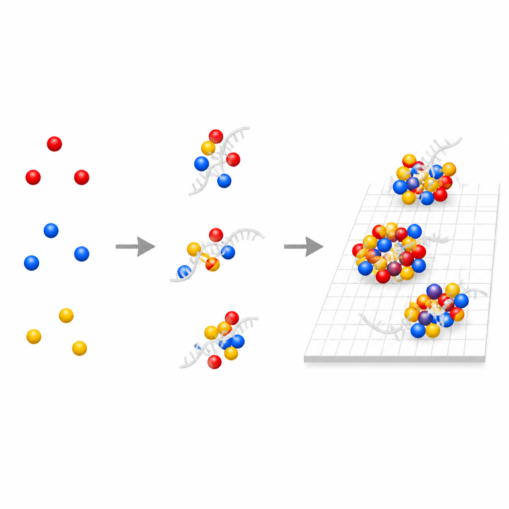
The researchers build their test around metal nanoparticles that shine in different colors under special lighting. Gold–coated silica particles appear red, while silver and small gold particles look blue or green. When a viral genetic strand or a piece of DNA is present, it can bind to DNA “handles” on these particles and pull them together into tiny clusters. These clusters scatter light differently from single particles. Instead of relying on a big change in color from the whole sample, the team focuses on the detailed patterns of color and shape produced by thousands of individual particles.
Simple chip and low-cost imaging
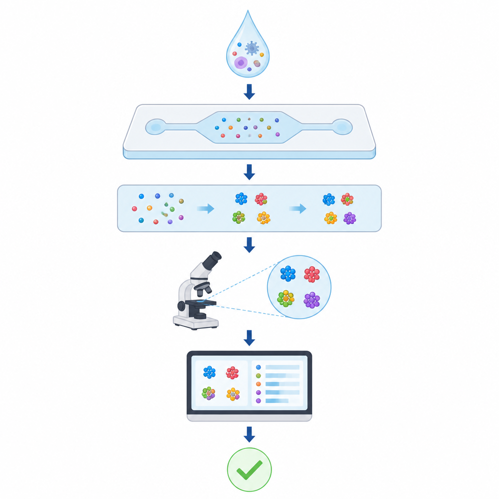
To keep the chemistry and hardware simple, the authors design a one-pot assay that does not need enzymes, amplification, or purification. For synthetic DNA targets, they mix the nanoparticles with carefully chosen buffers and heat briefly so DNA strands can bridge the particles. For real SARS-CoV-2 virus, they add strong detergents and a protein-digesting enzyme that both break open viral shells and protect the genetic material, all in the same tube. The mixture is then loaded into a thin chamber on a coverslip and imaged with a home-built dark-field microscope that uses inexpensive light-emitting diodes and a standard color camera to record the scattered light from many small regions.
Teaching a neural network to read particle patterns
Each microscope image contains up to several thousand bright spots, each corresponding to either a single nanoparticle or a small cluster. The team first cleans and normalizes these images so that variations in lighting and focus are reduced. They then cut out tiny image patches around each spot and feed them into a custom deep learning model called Mc-GNN. This model does two key things: it uses special filters that emphasize different shapes, such as circles, rings, edges, or square-like patterns, and it treats the outputs of these filters as nodes in a small graph so it can learn how different shape cues relate to each other. By learning these relationships across all particles in an image, the model estimates which concentration of viral material or synthetic DNA was present in the sample.
Performance compared with other methods
The authors rigorously compare their approach to more traditional machine learning models that rely on average color and simple shape measures from each image. Those methods reach around ninety percent accuracy at best and struggle to distinguish low target levels from controls, especially for whole-virus samples. Off-the-shelf deep learning models that look at particles one by one or treat the image as a whole also fall short. In contrast, Mc-GNN achieves very high recall scores across all tested concentrations, including femtomolar levels of synthetic DNA and viral RNA. It processes thousands of particle patches in a single pass while using less than two gigabytes of graphics memory, meaning it can run quickly on a consumer-grade graphics card.
What this could mean for future testing
By shifting much of the complexity from the chemistry and optics into software, this work outlines a path toward portable tests that are both sensitive and affordable. The assay still does not match the sensitivity of the most advanced laboratory genetic tests, but it already reaches virus levels relevant for real infections without requiring amplification or costly reagents. Because the method reads general patterns of nanoparticle clustering, it should be adaptable to new targets and could be combined with compact or smartphone-based microscopes. With further validation on real patient samples and refined hardware, such morphology-guided deep learning could help bring trustworthy molecular diagnostics closer to clinics, workplaces, and homes.
Citation: Jhawar, K., Chu, XL., DeGrandchamp, J.B. et al. Morphology-guided deep learning for nanoparticle agglomeration diagnostic assays. Sci Rep 16, 15248 (2026). https://doi.org/10.1038/s41598-026-45423-2
Keywords: nanoparticle diagnostics, deep learning assay, SARS-CoV-2 detection, dark-field microscopy, graph neural network